J Korean Ophthalmol Soc.
2014 Mar;55(3):443-448. 10.3341/jkos.2014.55.3.443.
Two Cases of Superior Limbic Keratoconjunctivitis Treated with Bevacizumab and Triamcinolone Injection
- Affiliations
-
- 1Department of Ophthalmology, Chung-Ang University College of Medicine, Seoul, Korea. jck50ey@kornet.net
- KMID: 2218277
- DOI: http://doi.org/10.3341/jkos.2014.55.3.443
Abstract
- PURPOSE
To report two cases of intractable superior limbic keratoconjunctivitis (SLK) treated with bevacizumab and triamcinolone injection.
CASE SUMMARY
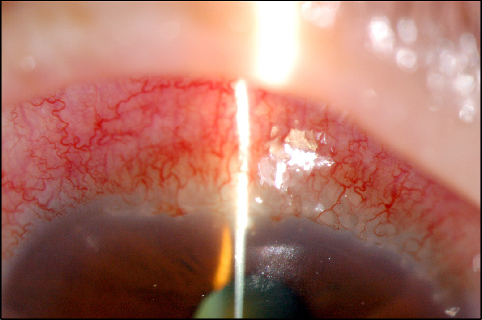
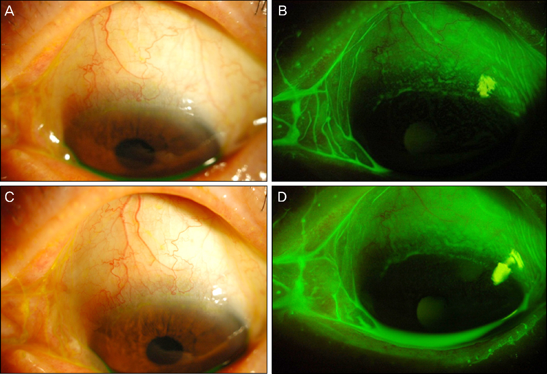
A 69-year-old female visited our clinic with pain in the left eye for 3 days and was diagnosed with SLK in her left eye. After 3 months of using steroid eye drops, artificial tears, and oral steroid intermittently, there was no improvement in symptoms and signs, thus this case was considered intractable with the conventional therapy. A mixture of bevacizumab (0.15 cc) and triamcinolone (0.05 cc) was injected into the sub-tenon's capsule of the left eye. After 1 week, all symptoms and signs disappeared, and there was no recurrence for 6 months. A 55-year-old female was transferred to our clinic due to SLK that did not respond to artificial tears, steroid eye drops, punctal occlusion, and botox injection for 3 months. A mixture of bevacizumab (0.15 cc) and triamcinolone (0.05 cc) was injected into the sub-tenon's capsule of the left eye. After 2 weeks, all symptoms and signs were improved, and there was no recurrence for 4 months.
CONCLUSIONS
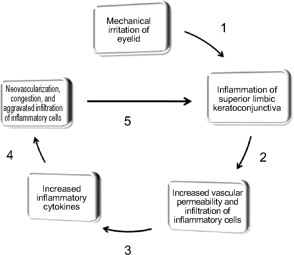
The presented 2 SLK cases are meaningful, because neovascularization disappeared and controlled inflammation was obtained following sub-tenon injection with both bevacizumab and triamcinolone.
MeSH Terms
Figure
Reference
-
References
1. Nelson JD. Superior limbic keratoconjunctivitis (SLK). Eye (Londo). 1989; 3:180–9.
Article2. Confino J, Brown SI. Treatment of superior limbic keratoconjunctivitis with topical cromolyn sodium. Ann Ophthalmol. 1987; 19:129–31.3. Sahin A, Bozkurt B, Irekec M. Topical cyclosporine a in the treatment of superior limbic keratoconjunctivitis: a long-term fol-low-up. Cornea. 2008; 27:193–5.4. Chun YS, Kim JC. Treatment of superior limbic keratoconjunctivitis with a large-diameter contact lens and Botulium Toxin A. Cornea. 2009; 28:752–8.5. Shen YC, Wang CY, Tsai HY, Lee YF. Supratarsal triamcinolone injection in the treatment of superior limbic keratoconjunctivitis. Cornea. 2007; 26:423–6.
Article6. Yang HY, Fujishima H, Toda I, et al. Lacrimal punctal occlusion for the treatment of superior limbic keratoconjunctivitis. Am J Ophthalmol. 1997; 124:80–7.
Article7. Sun YC, Hsiao CH, Chen WL, et al. Conjunctival resection combined with tenon layer excision and the involvement of mast cells in superior limbic keratoconjunctivitis. Am J Ophthalmol. 2008; 145:445–52.
Article8. Udell IJ, Kenyon KR, Sawa M, Dohiman CH. Treatment of superior limbic keratoconjunctivitis by thermocauterization of the superior bulbar conjunctiva. Ophthalmol. 1986; 93:162–6.
Article9. Kojima T, Matsumoto Y, Ibrahim OM, et al. In vivo evaluation of superior limbic keratoconjunctivitis using laser scanning confocal microscopy and conjunctival impression cytology. Invest Ophthalmol Vis Sci. 2010; 51:3986–92.
Article10. Kadrmas EF, Bartley GB. Superior limbic keratoconjunctivitis. A prognostic sign for severe Gravesophthalmopathy. Ophthalmol. 1995; 102:1472–5.11. Matsuda A, Tagawa Y, Matsuda H. TGF-beta2, tenascin, and in-tegrin beta1 expression in superior limbic keratoconjunctivitis. Jpn J Ophthalmo. 1999; 43:251–6.12. Cher I. Superior limbic keratoconjunctivitis: multifactorial me-chanical pathogenesis. Clin Experiment Ophthalmol. 2000; 28:181–4.
Article13. Metcalfe DD, Baram D, Mekori YA. Mast cells. Physiol Rev. 1997; 77:1033–79.
Article14. Kojima T, Matsumoto Y, Ibrahim OM, et al. In vivo evaluation of superior limbic keratoconjunctivitis using laser scanning confocal microscopy and conjunctival impression cytology. Invest Ophthalmol Vis Sci. 2010; 51:3986–92.
Article15. Jackson JR, Seed MP, Kircher CH, et al. The codependence of an-giogenesis and chronic inflammation. FASEB J. 1997; 11:457–65.
Article16. Ferrara N, Hillan KJ, Gerber HP, Novotny W. Discovery and development of bevacizumab, an anti-VEGF antibody for treating cancer. Nat Rev Drug Discov. 2004; 3:391–400.
Article17. Nakao S, Zandi S, Lara-Castillo N, et al. Larger therapeutic win-dow for steroid versus VEGF-A inhibitor in inflammatory angio-genesis: surprisingly similar impact on leukocyte infiltration. Invest Ophthalmol Vis Sci. 2012; 53:3296–302.
Article18. Oh JY, Kim MK, Shin MS, et al. The anti-inflammatory effect of subconjunctival bevacizumab on chemically burned rat corneas. Curr Eye Res. 2009; 34:85–91.
Article19. Wershil BK, Furuta GT, Lavigne JA, et al. Dexamethasone and cyclosporin A suppress mast cell-leukocyte cytokine cascades by multiple echanisms. Int Arch Allergy Immunol. 1995; 107:323–4.20. Itakura H, Akiyama H, Hagimura N, et al. Triamcinolone acetonide suppresses interleukin-1 beta-mediated increase in vascular endothelial growth factor expression in cultured rat Muller cells. Graefes Arch Clin Exp Ophthalmol. 2006; 244:226–31.21. Ferrante P, Ramsey A, Bunce C, Lightman S. Clinical trial to compare efficacy and side-effects of injection of posterior sub-Tenon triaet al. mcinolone versus orbital floor methylprednisolone in the management of posterior uveitis. Clin Exp Ophthalmol. 2004; 32:563–8.22. You IC, Kang IS, Lee SH, Yoon KC. Therapeutic effect of subconjunctival injection of bevacizumab in the treatment of corneal neovascularization. Acta Ophthalmol. 2009; 87:653–8.
Article
- Full Text Links
- Actions
-
Cited
- CITED
-
- Close
- Share
- Similar articles
-
- Superior Limbic Keratoconjunctivits
- A Case of Conjunctival Lithiasis with Clinical Manifestations of Superior Limbic Keratoconjunctivitis
- A Case of Conjunctival Lymphangioma With Clinical Manifestations of Superior Limbic Keratoconjunctivitis After Upper Lid Blepharoplasty
- The Effect of Intravitreal Triamcinolone Acetonide and Bevacizumab Injection on Intraocular Pressure
- Comparison of Intravitreal Bevacizumab Alone Injection and Intravitreal Combination Low-Dose Bevacizumab-Triamcinolone Injection or Diabetic Macular Edema


