A Case of Uneventful Cataract Surgery in Idiopathic True Exfoliation Patient
- Affiliations
-
- 1Department of Ophthalmology and The Institute of Vision Research, Yonsei University College of Medicine, Seoul, Korea.
- 2Siloam Eye Hospital, Seoul, Korea. 24mini@naver.com
- KMID: 2218101
- DOI: http://doi.org/10.3341/jkos.2014.55.5.766
Abstract
- PURPOSE
We present a case of uneventful cataract surgery in an idiopathic true exfoliation patient with areas of capsular delamination based on scanning electron microscope and transmission electron microscope results.
CASE SUMMARY
A 77-year-old male presented with gradual deterioration of vision over 1 year in duration. Slit lamp examination revealed bilateral nuclear sclerotic cataracts with ring-shaped fibrous membrane floating within the anterior chamber in the right eye. In addition, the patient was diagnosed with cataract and true exfoliation of the right eye. He underwent uneventful phacoemulsification and posterior chamber intraocular lens implantation by placing capsulorrhexis outside the delaminated capsule margin. At 6 months after cataract surgery, the patient showed favorable visual outcome with uncorrected vision of 20/20 and intraocular pressure of 18 mm Hg in the right eye.
MeSH Terms
Figure
-

Figure 1. Slit lamp examination showing floating ring-shaped fibrous membrane (arrows) in the anterior chamber in the right eye (A) before dilation, (B) after dilation of the pupil, and (C) with retroillumination.
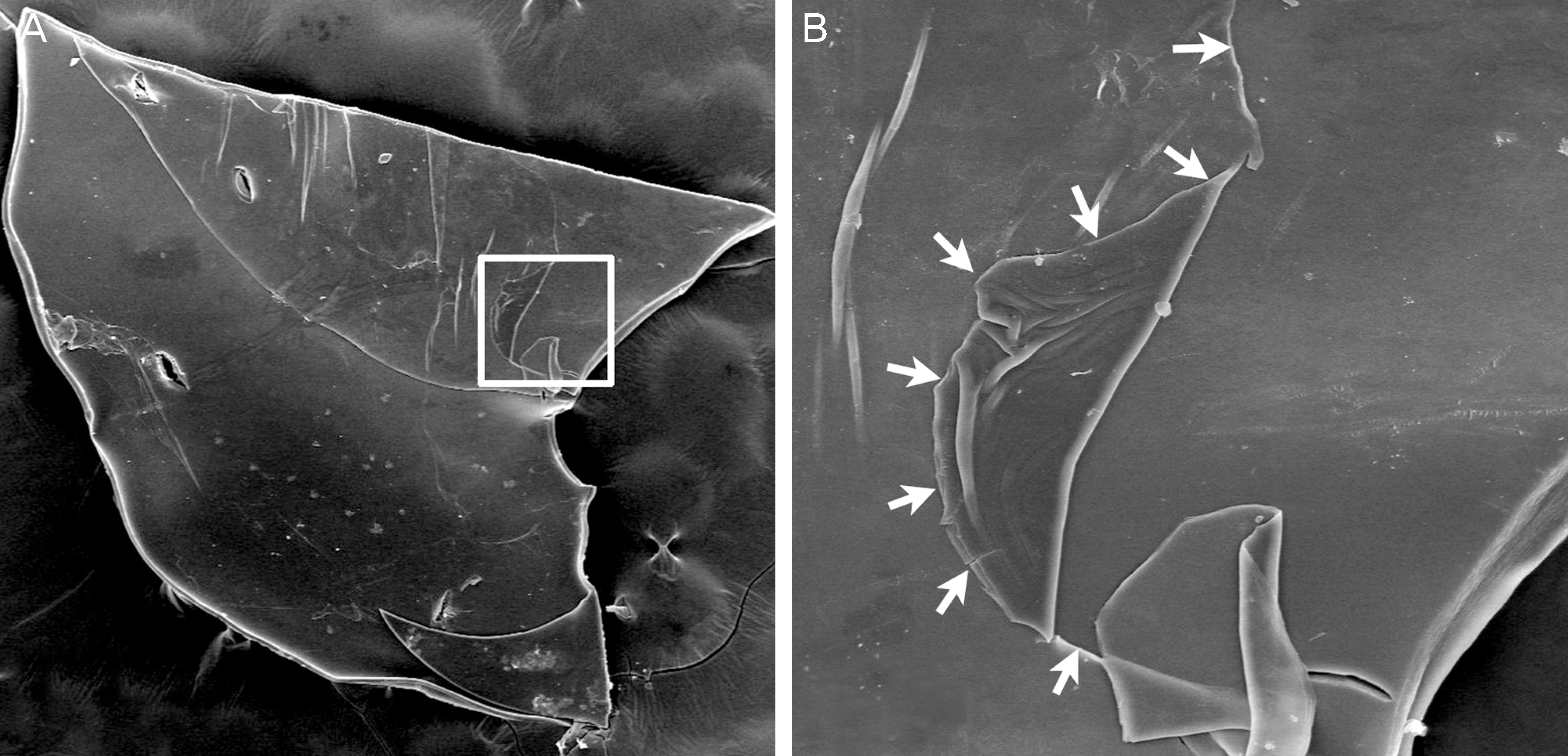
Figure 2. Scanning electron micrographs of the anterior lens capsule after capsulorrhexis at different magnifications. (A) At low magnification (x50). Part of the area of delamination indicated with white rectangle was re-examined at higher magnification. (B) At high magnification (x300). The arrows show the exfoliation flap.
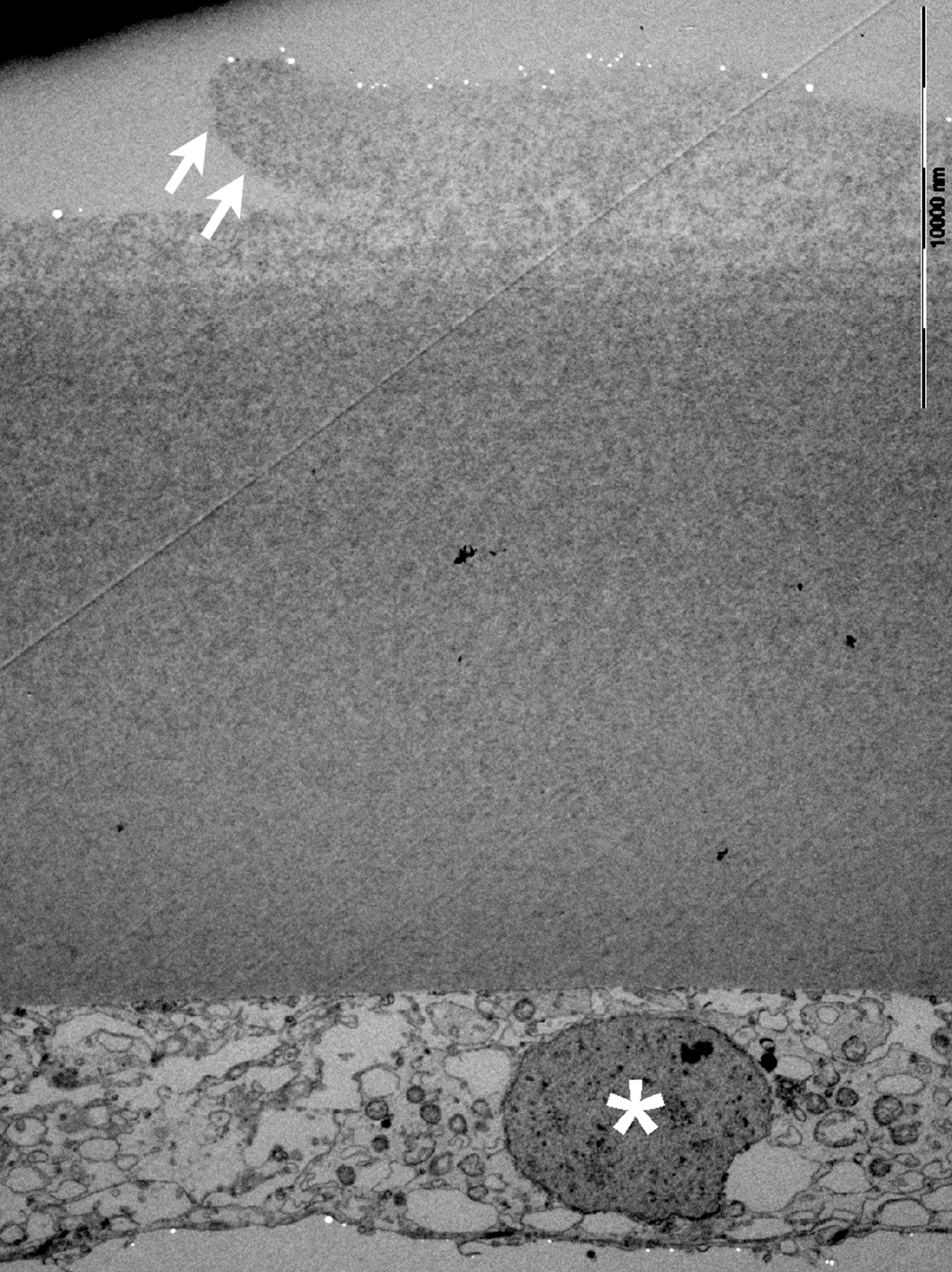
Figure 3. Transmission electron micrograph of anterior lens capsule after capsulorrhexis showing the superficial layer of the anterior lens capsule (arrows) separated from the deep layer and lens epithelium (asterisk).
Reference
-
References
1. Oharazawa H, Suzuki H, Matsui H, et al. Two cases of true exfoliation of the lens capsule after cataract surgery. J Nippon Med Sch. 2007; 74:55–60.
Article2. Elshnig . Detachment of the zonular lamella in glassblowers. Klin Monatsbl Augenheilkd. 1922; 69:732–4.3. Lee SH, Lee JE. True exfoliation of the lens capsule. J Korean Ophthalmol Soc. 2001; 42:392–5.4. Kim KH, Chung ES, Chung TY. Radial extension of capsulorhexis in true exfoliation patient: a potentially hazardous complication. J Cataract Refract Surg. 2009; 35:590–2.
Article5. Yamamoto Y, Nakakuki T, Nishino K, et al. Histological and clin-ical study of eyes with true exfoliation and a double-ring sign on the anterior lens capsule. Can J Ophthalmol. 2009; 44:657–62.
Article6. Burde RM, Bresnick G, Uhrhammer J. True exfoliation of the lens cpsule. An electron microscop study. Arch Ophthalmol. 1969; 82:651–3.7. Callahan A, Klien BA. Thermal detachment of the anterior lamella of the anterior lens capsule; a clinical and histopathologic study. AMA Arch Ophthalmol. 1958; 59:73–80.8. Yamamoto N, Miyagawa A. True exfoliation of the lens capsule following uveitis. Graefes Arch Clin Exp Ophthalmol. 2000; 238:1009–10.
Article9. Cashwell LF Jr, Holleman IL, Weaver RG, van Rens GH. Idiopathic true exfoliation of the lens capsule. Ophthalmology. 1989; 96:348–51.
Article10. Dvorak-Theobald G. Pseudoexfoliation of the lens capsule: relation to true exfoliation of the lens capsule as reported in the liter-ature, and role in the production of glaucoma capsulocuticulare. Trans Am Ophthalmol Soc. 1953; 51:385–407.11. Fiore PM, Shingleton BJ. Senile lens exfoliation. JAMA. 1990; 264:2755.
Article12. Braude LS, Edward DP. Partial splitting of the anterior lens capsule giving a ‘double-ring’ sign. Arch Ophthalmol. 1995; 113:705–8.
Article13. Wollensak G, Wollensak J. Double contour of the lens capsule edges after continuous curvilinear capsulorhexis. Graefes Arch Clin Exp Ophthalmol. 1997; 235:204–7.
Article14. Ataka S, Kohno T, Kurita K, et al. Histopathological study of the anterior lens capsule with a double-ring sign. Graefes Arch Clin Exp Ophthalmol. 2004; 242:245–9.
Article15. Tan DK, Aung T, Perera SA. Novel method of assessing de-lamination of the anterior lens capsule using spectral-domain opti-cal coherence tomography. Clin Ophthalmol. 2012; 6:945–8.
Article16. Kulkarni AR, Al-Ibrahim J, Haider S, et al. Phacoemulsification in true exfoliation of the lens capsule: a case series. Eye (Lond). 2007; 21:835–7.
Article17. Rossiter J, Morris A. Trypan blue vital staining of the anterior lens capsule in the management of cataract in true exfoliation of the lens capsule. Eye (Lond). 2005; 19:809–10.
Article
- Full Text Links
-
- Actions
-
Cited
- CITED
-
- Close
- Share
-
- Similar articles
-
- True Exfoliation of the Lens Capsule
- Lens Dislocation during Extracapsular Cataract Extraction in Exfoliation Syndrome: A Case Report
- A Histopathological Study on the Production of Exfoliation Material in Eyes with Exfoliation Syndrome
- A Histopathologic Study of the Iris in a Patient with Exfoliation Syndrome
- Refractive Outcomes of Uneventful Cataract Surgery in Pseudoexfoliation Syndrome and Pseudoexfoliation Glaucoma
