J Korean Ophthalmol Soc.
2013 Nov;54(11):1715-1722. 10.3341/jkos.2013.54.11.1715.
Correlation Between Optical Coherence Tomography and Visual-Acuity in Chronic Central Serous Chorioretinopathy Treated with Half Dose Photodynamic Therapy
- Affiliations
-
- 1The Institute of Vision Research, Department of Ophthalmology, Yonsei University College of Medicine, Seoul, Korea.
- 2Siloam Eye Hospital, Seoul, Korea. kym1248@gmail.com
- KMID: 2217919
- DOI: http://doi.org/10.3341/jkos.2013.54.11.1715
Abstract
- PURPOSE
To evaluate the results of half-dose photodynamic therapy (PDT) and determine the correlation between morphological changes measured by spectral-domain optical coherence tomography (SD-OCT) and visual acuity in patients with chronic central serous chorioretinopathy (CSC).
METHODS
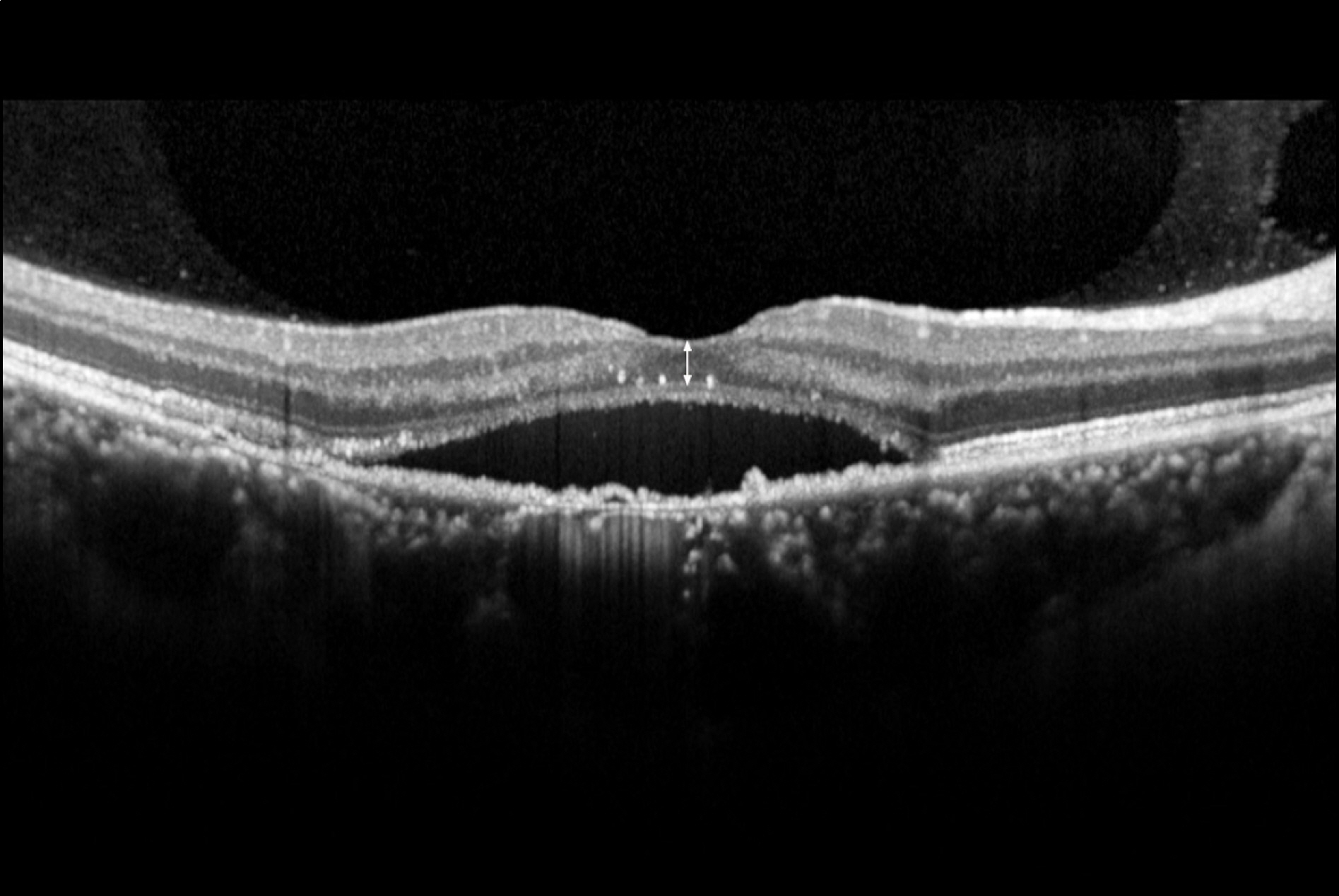
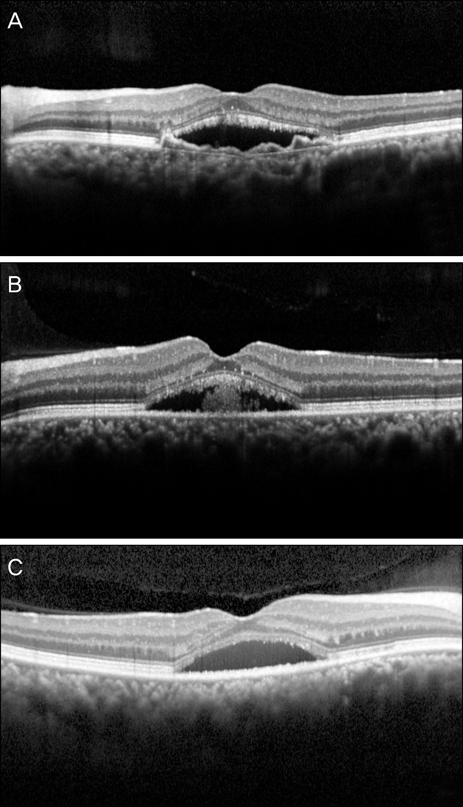
Twenty-five eyes of 25 patients with chronic CSC who had received half-dose verteporfin PDT were enrolled in the present study. The best-corrected visual acuity (BCVA), outer nuclear layer (ONL) thickness, and the integrity of the photoreceptor inner and outer segment junction (IS/OS) using SD-OCT were evaluated at baseline and at 3 and 6 months after treatment.
RESULTS
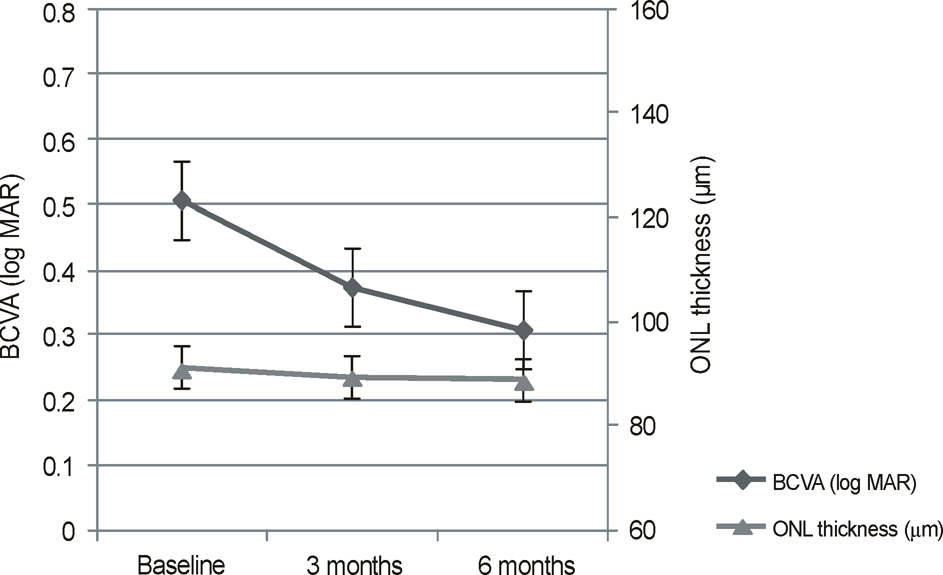
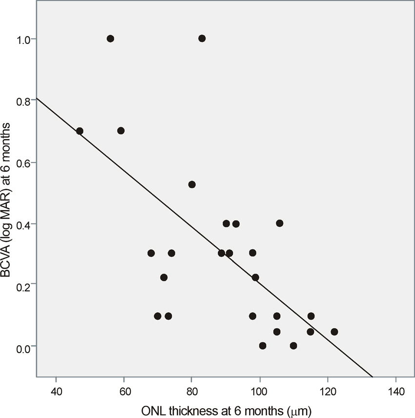
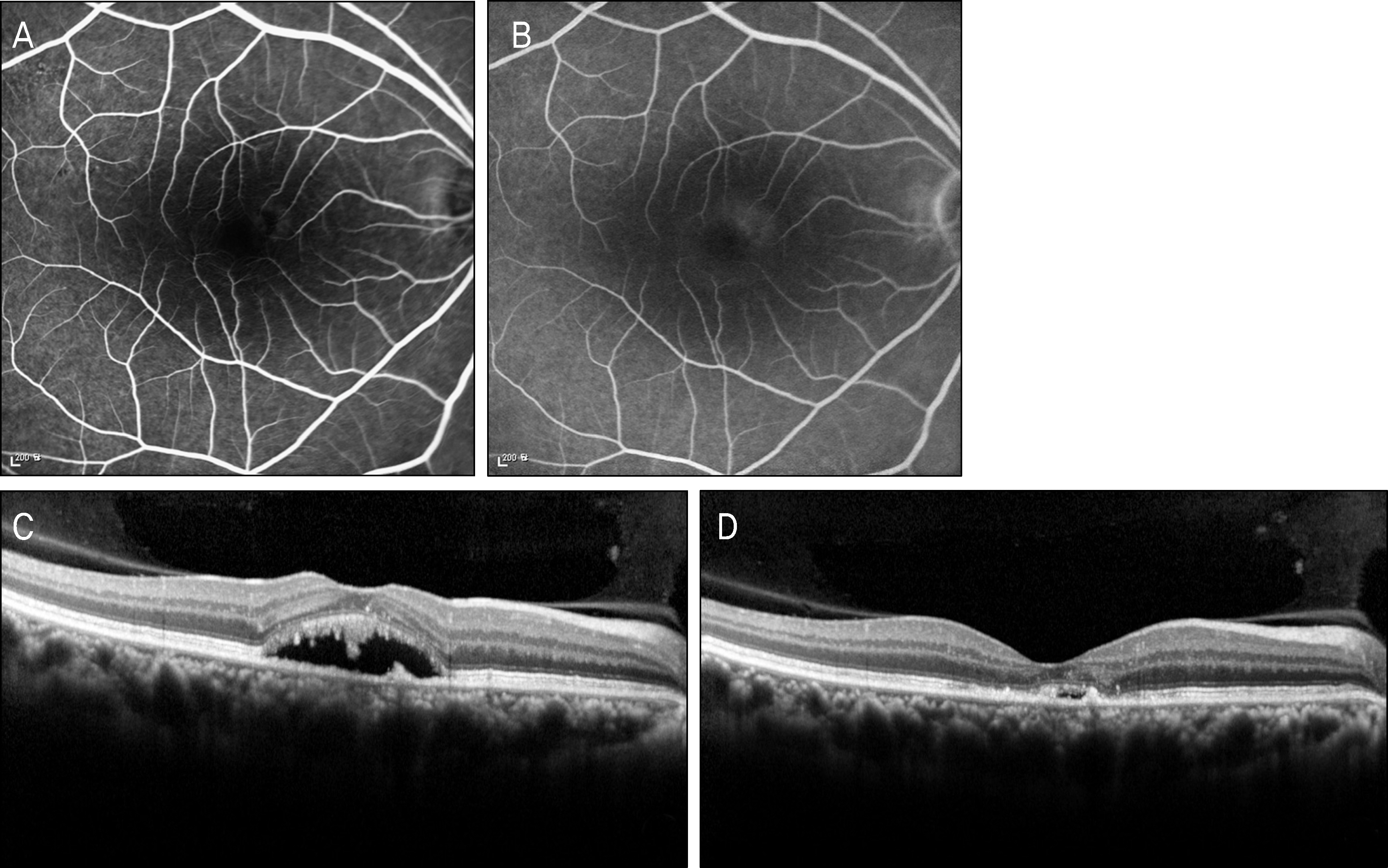
The neurosensory retinal detachment disappeared in all eyes 6 months after treatment. The BCVA improved significantly from 0.50 +/- 0.32 to 0.31 +/- 0.29 log MAR at 6 months (p < 0.001). The average ONL thickness at the central fovea was 88.76 +/- 19.95 microm at 6 months and the ONL thickness was well correlated with the BCVA (gamma = -0.64; p = 0.001). There was no significant correlation between the status of IS/OS and the BCVA.
CONCLUSIONS
Half-dose PDT is effective in treating chronic CSC resulting in visual improvement and complete resolution of neurosensory retinal detachment. The ONL thickness which was positively correlated with the BCVA could be an indicator for visual prognosis of chronic CSC.
Keyword
MeSH Terms
Figure
Reference
-
References
1. Gass JD. Pathogenesis of disciform detachment of the neuroepithelium. Am J Ophthalmol. 1967; 63:1–139.2. Yap EY, Robertson DM. The long-term outcome of central serous chorioretinopathy. Arch Ophthalmol. 1996; 114:689–92.
Article3. Wang MS, Sander B, Larsen M. Retinal atrophy in idiopathic cen- tral serous chorioretinopathy. Am J Ophthalmol. 2002; 133:787–93.4. Loo RH, Scott IU, Flynn HW Jr, et al. Factors associated with re- duced visual acuity during long-term follow-up of patients with idiopathic central serous chorioretinopathy. Retina. 2002; 22:19–24.5. Robertson DM. Argon laser photocoagulation treatment in central serous chorioretinopathy. Ophthalmology. 1986; 93:972–4.
Article6. Piccolino FC. Laser treatment of eccentric leaks in central serous chorioretinopathy resulting in disappearance of untreated juxtafo- veal leaks. Retina. 1992; 12:96–102.7. Colucciello M. Choroidal neovascularization complicating photo- dynamic therapy for central serous retinopathy. Retina. 2006; 26:239–42.8. Tzekov R, Lin T, Zhang KM, et al. Ocular changes after photo-dynamic therapy. Invest Ophthalmol Vis Sci. 2006; 47:377–85.
Article9. Lai TY, Chan WM, Lam DS. Transient reduction in retinal function revealed by multifocal electroretinogram after photodynamic therapy. Am J Ophthalmol. 2004; 137:826–33.
Article10. Kim JL, Kim HW, Yoon IH. Photodynamic therapy with vertepofin for short time for chronic central serous chorioretinopathy. J Korean Ophthalmol Soc. 2008; 49:1078–86.
Article11. Chang MH, Kim SW, Oh JR, Huh K. Photodynamic therapy with verteporfin using half fluence for chronic central serous chorioretinopathy. J Korean Ophthalmol Soc. 2009; 50:1326–33.
Article12. Rouvas A, Stavrakas P, Theodossiadis PG, et al. Long-term results of half-fluence photodynamic therapy for chronic central serous chorioretinopathy. Eur J Ophthalmol. 2012; 22:417–22.
Article13. Smretschnig E, Ansari-Shahrezaei S, Moussa S, et al. Half-fluence photodynamic therapy in acute central serous chorioretinopathy. Retina. 2012; 32:2014–9.
Article14. Bressler NM. Treatment of age-related macular degeneration with photodynamic therapy (TAP) Study Group. Photodynamic therapy of subfoveal choroidal neovascularization in age-related macular degeneration with verteporfin: one-year results of 2 randomized clinical trials-TAP report 1. Arch Ophthalmol. 1999; 117:1329–45.15. Bressler NM. Treatment of age-related macular degeneration with photodynamic therapy (TAP) Study Group. Photodynamic therapy of subfoveal choroidal neovascularization in age-related macular degeneration with verteporfin: two-year results of 2 randomized clinical trials - TAP report 2. Arch Ophthalmol. 2001; 119:198–207.16. Piccolino FC, Eandi CM, Ventre L, et al. Photodynamic therapy for chronic central serous chorioretinopathy. Retina. 2003; 23:752–63.
Article17. Yannuzzi LA, Slakter JS, Gross NE, et al. Indocyanine green an- giography-guided photodynamic therapy for treatment of chronic central serous chorioretinopathy: a pilot study. Retina. 2003; 23:288–98.18. Taban M, Boyer DS, Thomas EL, et al. Chronic central serous cho- rioretinopathy: photodynamic therapy. Am J Ophthalmol. 2004; 137:1073–80.19. Uetani R, Ito Y, Oiwa K, et al. Half-dose vs one-third-dose photo- dynamic therapy for chronic central serous chorioretinopathy. Eye. 2012; 26:640–9.20. Fujita K, Shinoda K, Imamura Y, et al. Correlation of integrity of cone outer segment tips line with retinal sensitivity after half-dose photodynamic therapy for chronic central serous chorioretinopathy. Am J Ophthalmol. 2012; 154:579–85.
Article21. Matsumoto H, Sato T, Kishi S. Outer nuclear layer thickness at the fovea determines visual outcomes in resolved central serous chorioretinopathy. Am J Ophthalmol. 2009; 148:105–10e1.
Article22. Hata M, Oishi A, Shimozono M, et al. Early changes in foveal thickness in eyes with central serous chorioretinopathy. Retina. 2013; 33:296–301.
Article23. Iida T, Yannuzzi LA, Spaide RF, et al. Cystoid macular degener- ation in chronic central serous chorioretinopathy. Retina. 2003; 23:1–7.24. Eandi CM, Chung JE, Cardillo-Piccolino F, et al. Optical coher- ence tomography in unilateral resolved central serous choriore- tinopathy. Retina. 2005; 25:417–21.25. Matsumoto H, Kishi S, Otani T, et al. Elongation of photoreceptor outer segment in central serous chorioretinopathy. Am J Ophthalmol. 2008; 145:162–8e1.
Article26. Ojima Y, Tsujikawa A, Yamashiro K, et al. Restoration of outer segments of foveal photoreceptors after resolution of central se- rous chorioretinopathy. Jpn J Ophthalmol. 2010; 54:55–60.
- Full Text Links
- Actions
-
Cited
- CITED
-
- Close
- Share
- Similar articles
-
- Photodynamic Therapy with Vertepofin for Short Time for Chronic Central Serous Chorioretinopathy
- Photodynamic Therapy With Verteporfin using Half Fluence for Chronic Central Serous Chorioretinopathy
- The Use fulness of OCT[Optical Coherence Tomography]for the Diagnosis of Central Serous Choriore tinopathy
- Chronic Central Serous Chorioretinopathy: Photodynamic Therapy
- Measurement and Analysis of Serous Fluid in Central Serous Chorioretinopathy using OCT

