J Korean Ophthalmol Soc.
2014 Nov;55(11):1706-1709. 10.3341/jkos.2014.55.11.1706.
Treatment of Scleromalacia with Scleral Autograft
- Affiliations
-
- 1Department of Ophthalmology, Myongji Hospital, Goyang, Korea. eyeminerva@naver.com
- KMID: 2216622
- DOI: http://doi.org/10.3341/jkos.2014.55.11.1706
Abstract
- PURPOSE
To report a case of a patient who developed scleromalacia after cosmetic eye whitening conjunctivectomy and treated with scleral and conjunctival autograft.
CASE SUMMARY
A 42-year-old male patient who received cosmetic eye whitening conjunctivectomy in both eyes on the nasal side in 2008 developed scleromalacia in the left eye. Calcium deposits and deformed conjunctiva were removed from the left eye. Autogenous sclera and conjunctiva were obtained from the upper side of the left eye and autogenous graft was performed. Topical antibiotics, topical steroid, topical autologous serum, and antibiotic ointment were applied postoperatively. The patient was given oral steroid for 1 month after surgery. During the postoperative 6 months, the grafted autogenous sclera was well maintained and improved cosmetically.
CONCLUSIONS
In cases of scleromalacia occurring after cosmetic eye whitening conjunctivectomy, autogenous sclera can be considered as a treatment filler.
MeSH Terms
Figure
-
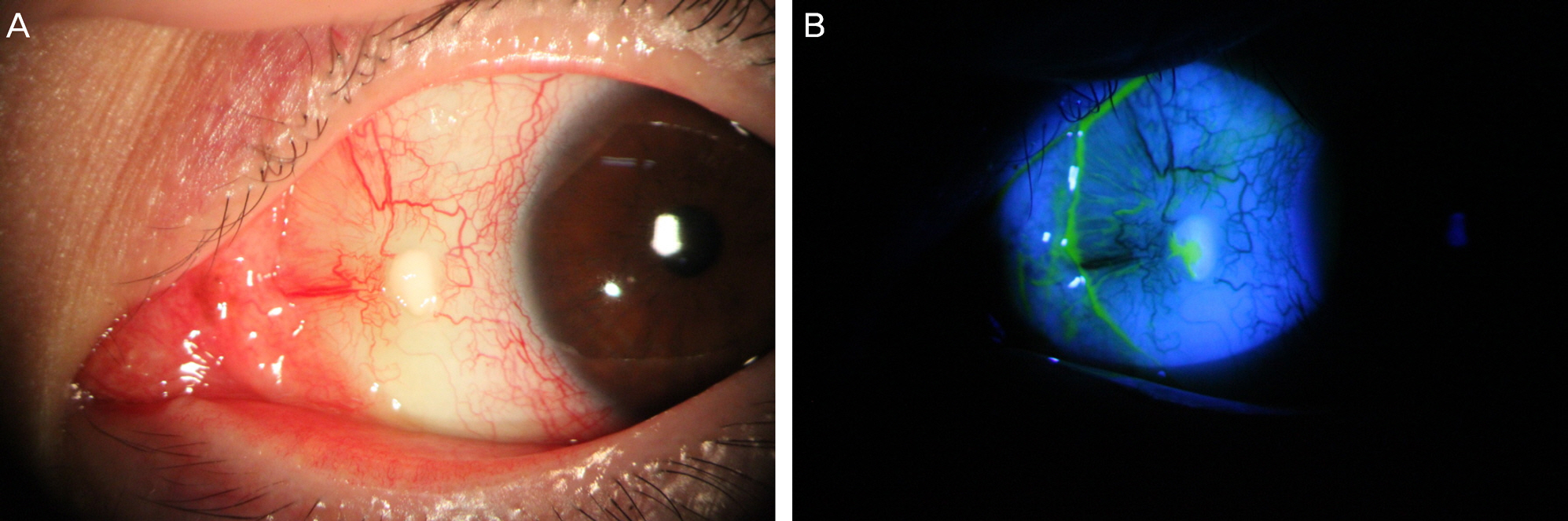
Figure 1. (A) Slit-lamp photographs of left eye show the thinned bare, avascular sclera, and (B) fluorescence stained lesion. White calcified plaque was noted on the nasal bulbar conjunctiva.
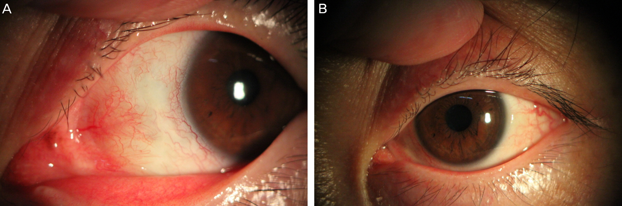
Figure 2. (A, B) Slit-lamp photographs of patient undergoing free conjunctival autograft with autogenous scleral graft for restoration of scleromalacia. Six months after surgery, scleral graft was well maintained and cosmetically improved.
Reference
-
References
1. Shin HY, Kim MS, Chung SK. The development of scleromalacia after regional conjunctivectomy with the postoperative application of mitomycin C as an adjuvant therapy. Korean J Ophthalmol. 2013; 27:208–10.
Article2. Dougherty PJ, Hardten DR, Lindstrom RL. Corneoscleral melt after pterygium surgery using a single intraoperative application of mitomycin-C. Cornea. 1996; 15:537–40.
Article3. Kwak JY, Chang HK. Autogenous temporalis fascia grafting and conjunctival flap transposition in scleromalacia after pterygium excision. J Korean Ophthalmol Soc. 2004; 45:180–6.4. Lee CO, Jong SH, Lee JJ. Autologous simple conjunctival graft and conjunctiva/tenon graft on focal scleromalacia. J Korean Ophthalmol Soc. 1997; 38:1737–41.5. Breslin CW, Katz JI, Kaufman HE. Surgical management of necrotizing scleritis. Arch Ophthalmol. 1977; 95:2038–40.
Article6. Song HY, Im JS, Kwak JY. Acellular dermal allograft transplantation in patients with scleromalacia after pterygium excision. J Korean Ophthalmol Soc. 2008; 49:1685–9.
Article7. Choi WS, Lee GJ, Park YJ, Lee KW. Scleral graft, free conjunctival autograft using tissue adhesive and temporary amniotic membrane transplantation in scleromalacia. J Korean Ophthalmol Soc. 2011; 52:1405–13.
Article8. Oh DH, Kim JC, Chun YS. A case of fusarium deep keratitis following scleral graft. J Korean Ophthalmol Soc. 2010; 51:606–10.
Article9. Kwon HJ, Nam SM, Lee SY, et al. Conjunctival flap surgery for calcified scleromalacia after cosmetic conjunctivectomy. Cornea. 2013; 32:821–5.
Article
- Full Text Links
-
- Actions
-
Cited
- CITED
-
- Close
- Share
-
- Similar articles
-
- Scleral Graft, Free Conjunctival Autograft Using Tissue Adhesive and Temporary Amniotic Membrane Transplantation in Scleromalacia
- Repair of scleromalacia with Preserved Scleral and Amniotic Membrane Transplantation
- Scleral Allografting and Amniotic Membrane Transplantation With Fibrin Glue in the Management of Scleromalacia
- Autogenous Temporalis Fascia Grafting and Conjunctival Flap Transposition in Scleromalacia after Pterygium Excision
- A Case of Scleromalacia Perforance That Developing after Surgery for Excision of the Pterygium in a Patient with Rheumatoid Arthritis
