Outer Retinal Tubulation in Chronic Central Serous Chorioretinopathy
- Affiliations
-
- 1Cheil Eye Hospital, Daegu, Korea. ppjinsun25@hanmail.net
- 2Department of Ophthalmology, Kyungpook National University School of Medicine, Daegu, Korea.
- KMID: 2216613
- DOI: http://doi.org/10.3341/jkos.2014.55.11.1642
Abstract
- PURPOSE
To evaluate outer retinal tubulation (ORT) found in chronic central serous chorioretinopathy (CSC) using color fundus photography and spectral domain optical coherence tomography (SD-OCT).
METHODS
ORT identified in patients with chronic CSC was examined using color fundus photography, fundus autofluorescence image, fluorescein angiography, indocyanin green angiography, and SD-OCT. The images were analyzed for morphological features, location, and size of ORT in the retinal layers.
RESULTS
ORT was detected in 3 of 342 (0.88%) chronic CSC patients. Color fundus photography revealed circular or ovoid shape with hollow lumen and deep yellowish border mainly appearing at the affected retinal pigment epithelium layer in the macular area. SD-OCT B-scan revealed hyperreflective material observed inside the hyporeflective internal space with hyperreflective border. ORT had circular or ovoid shape on the SD-OCT C-scan. ORT was primarily located on the outer nuclear layer in the retina, emanating to the inner nuclear layer and was not greater than 170 x 170 microm in size when measured with SD-OCT B-scan image.
CONCLUSIONS
ORT was identified in patients with chronic CSC which was observed using color fundus photography and circular or ovoid structure was observed using a SD-OCT C-scan.
Keyword
MeSH Terms
Figure
-
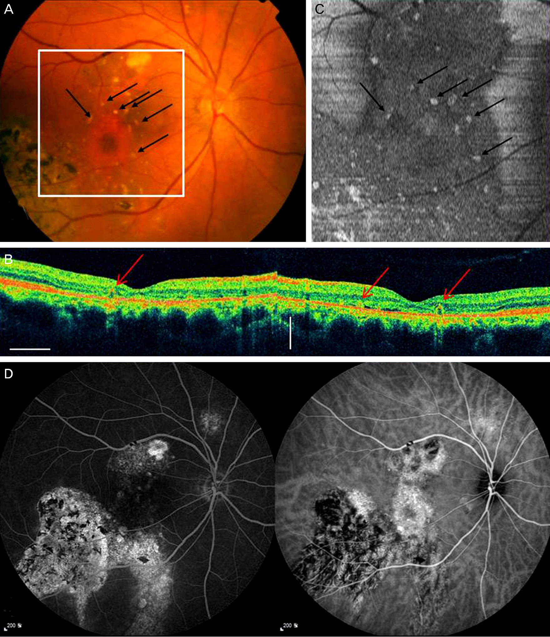
Figure 1. (A) Color fundus photography of right eye of a 54-years-old man with outer retinal tubulation in chronic central serous chorioretinopathy. Black arrows indicate some outer retinal tubulation which represents deep yellowish spots on color fundus photography. White box represents the area of B-scan and C-scan. (B) Spectral domain optical coherence tomography (SD-OCT) B-scan (left: horizontal section through the fovea, right: vertical section through the fovea) showed circular and ovoid structures with hyperreflective borders representing outer retinal tubulation in the outer nuclear layer (red arrows). (C) Spectral domain OCT C-scan shows multiple circular or ovoid shapes with hyperreflectivity, which correspond with deep yellowish spots on color fundus photography (black arrows). (D) Fluorescein angiography and indocyanin green angiography show retinal pigment epithelium track in chronic central serous chorioretinopathy.
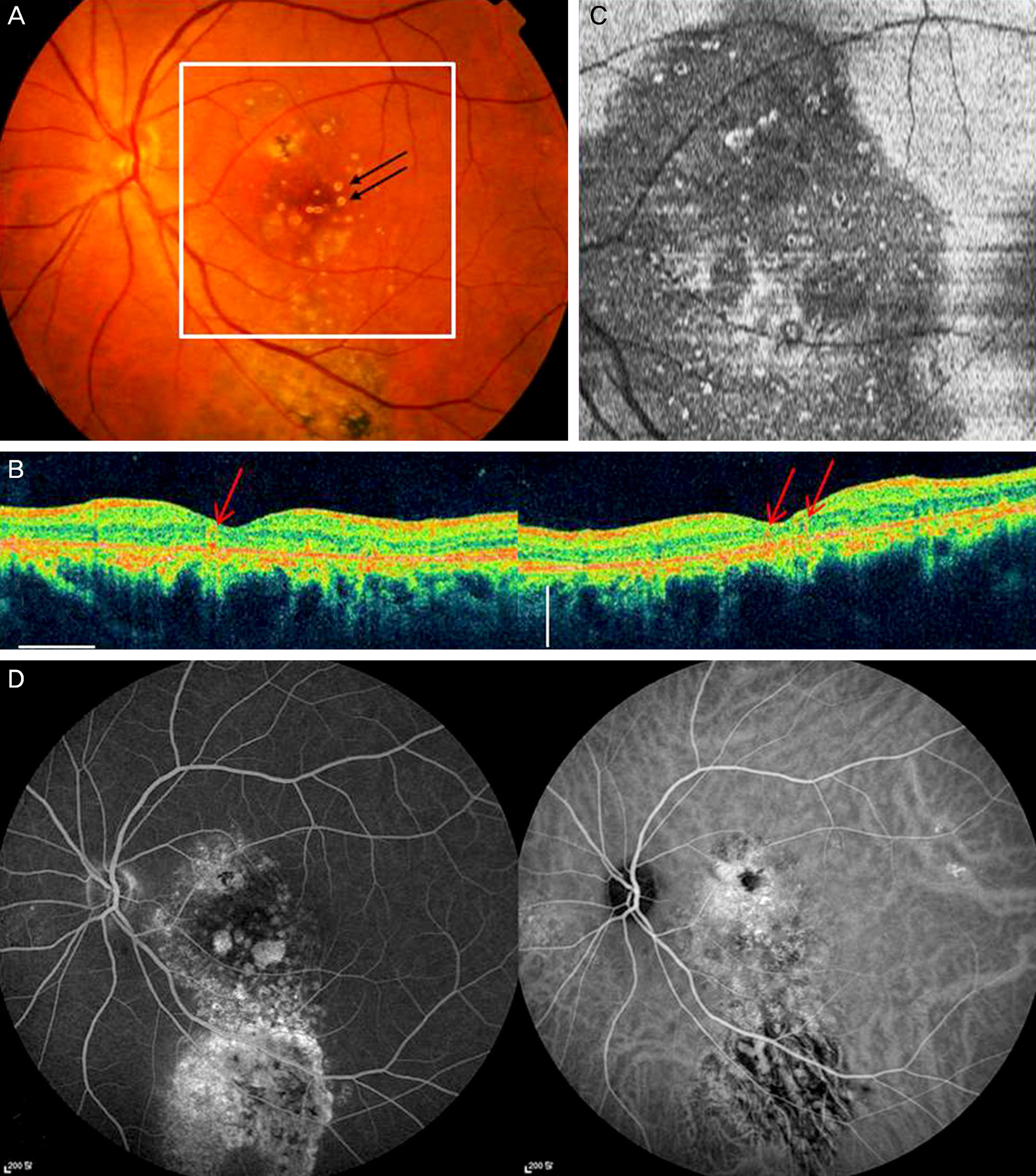
Figure 2. (A) Color fundus photography of left eye of a 54-years-old man with outer retinal tubulation in chronic central serous chorioretinopathy. Black arrows indicate outer retinal tubulation which correspond with red arrows in spectral domain OCT B-scan. White box represents the area of B-scan and C-scan. (B) Spectral domain optical coherence tomography (SD-OCT) B-scan (left: horizontal section through the fovea, right: vertical section through the fovea) showed circular and ovoid structures with hyperreflective borders representing outer retinal tubulation in the outer nuclear layer (red arrows). (C) Spectral domain OCT C-scan shows multiple circular or ovoid shapes with hyperreflectivity, which correspond with deep yellowish spots on color fundus photography. (D) Fluorescein angiography and indocyanin green angiography show retinal pigment epithelium track in chronic central serous chorioretinopathy.

Figure 3. (A) Color fundus photography of right eye of a 69-years-old man with outer retinal tubulation in chronic central serous chorioretinopathy. Black arrows indicate outer retinal tubulation which correspond with red arrows in spectral domain optical coherence tomography (SD-OCT) B-scan. White box represents the area of C-scan. (B) Spectral domain OCT B-scan showed circular and ovoid structures with hyperreflective borders and contained hyper-reflective material within their lumen representing outer retinal tubulation. (C) Spectral domain OCT C-scan showed circular pattern of outer retinal tubulation located near atrophic scar. (D) Fluorescein angiography and indocyanin green angiography show retinal pigment epithelium atrophy in chronic central serous chorioretinopathy.
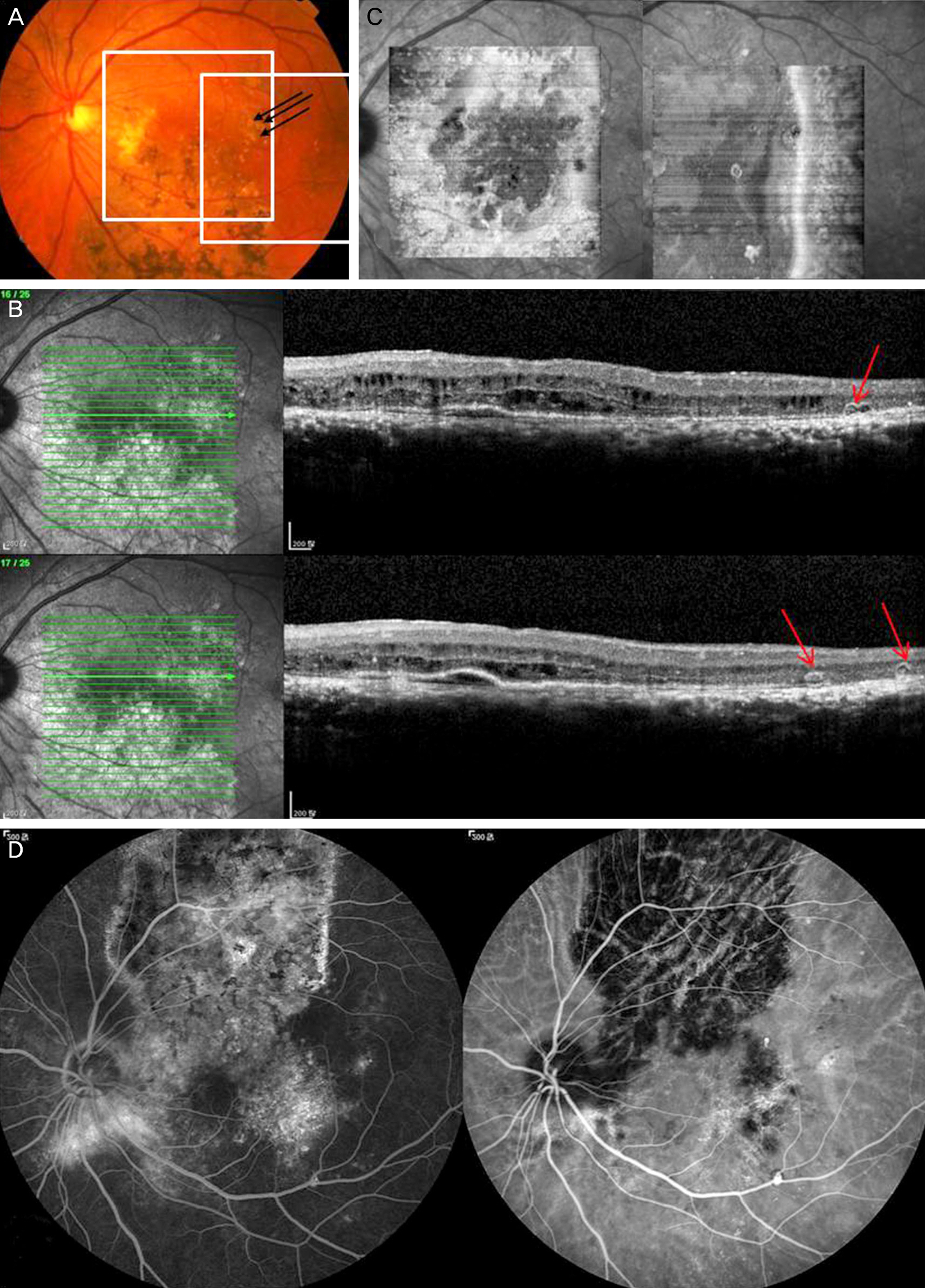
Figure 4. (A) Color fundus photography of left eye of a 69-years-old man with outer retinal tubulation in chronic central serous chorioretinopathy. Black arrows indicate outer retinal tubulation which correspond with red arrows in spectral domain optical coherence tomography (SD-OCT) B-scan. White box represents the area of C-scan. (B) Spectral domain OCT B-scan showed circular and ovoid structures with hyperreflective borders and contained hyperreflective material within their lumen representing outer retinal tubulation. (C) Spectral domain OCT C-scan showed circular pattern of outer retinal tubulation located near atrophic scar. (D) Fluorescein angiography and indocyanin green angiography show retinal pigment epithelium atrophy chronic central serous chorioretinopathy.
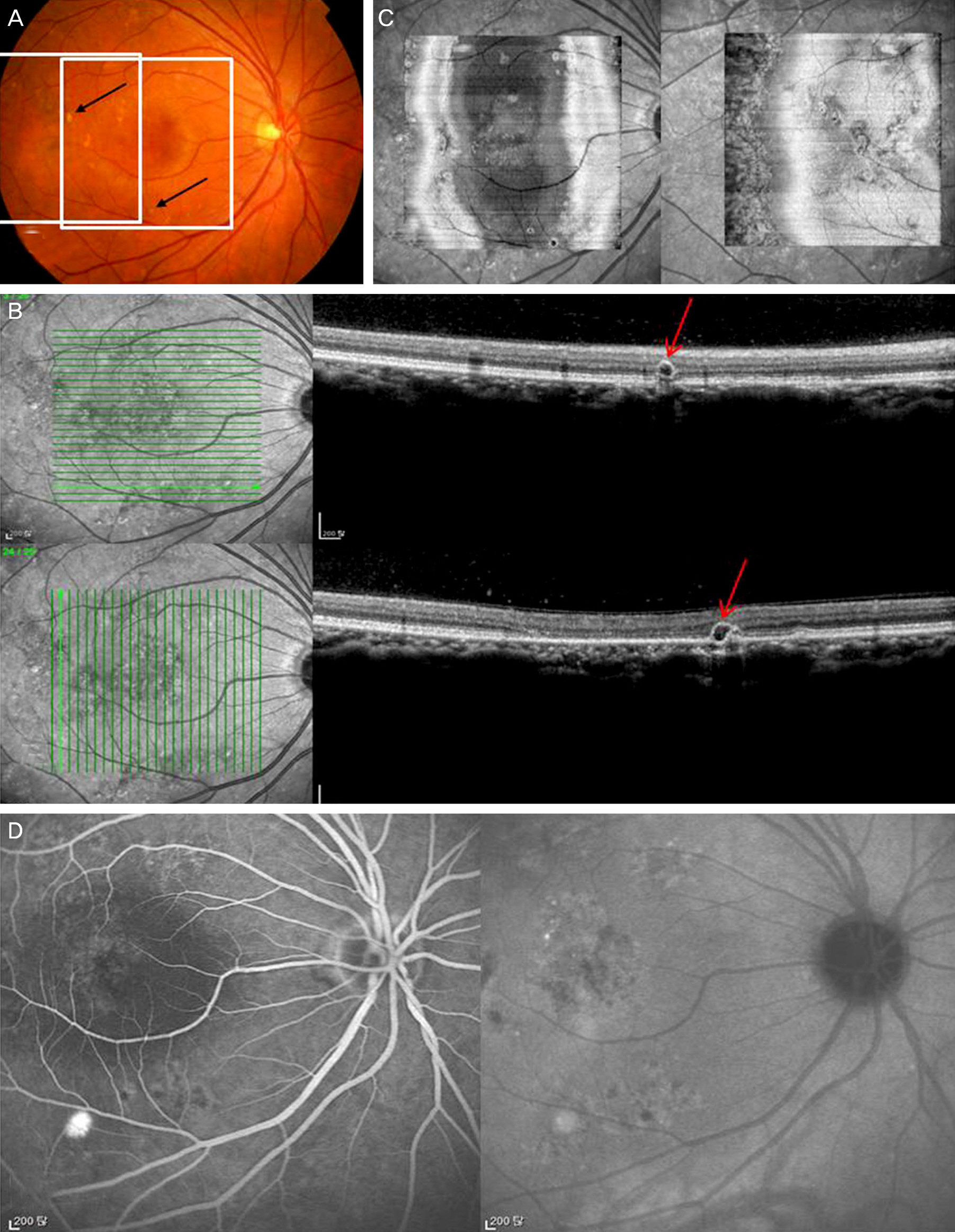
Figure 5. (A) Color fundus photography of right eye of a 57-years-old woman with outer retinal tubulation in chronic central serous chorioretinopathy. Black arrows indicate outer retinal tubulation which correspond with red arrows in spectral domain optical coherence tomography (SD-OCT) B-scan. White box represents the area of C-scan. (B) Spectral domain OCT B-scan showed circular and ovoid structures with hyperreflective borders representing outer retinal tubulation. (C) Spectral domain OCT C-scan showed circular pattern of outer retinal tubulation located near atrophic scar. (D) Fluorescein angiography and indocyanin green angiography show window defect in chronic central serous chorioretinopathy.
Reference
-
References
1. Zweifel SA, Engelbert M, Laud K, et al. Outer retinal tubulation: a novel optical coherence tomography finding. Arch Ophthalmol. 2009; 127:1596–602.2. Iriyama A, Aihara Y, Yanagi Y. Outer retinal tubulation in inherited retinal degenerative disease. Retina. 2013; 33:1462–5.
Article3. Yannuzzi LA. The retinal atlas. 1st ed.New York, NY, USA: Elsevier Health Sciences;2010. p. 158–60.4. Ellabban AA, Hangai M, Yamashiro K, et al. Tomographic fundus features in pseudoxanthoma elasticum: comparison with neovascular age-related macular degeneration in Japanese patients. Eye (Lond). 2012; 26:1086–94.
Article5. Sergouniotis PI, Davidson AE, Lenassi E, et al. Retinal structure, function, and molecular pathologic features in gyrate atrophy. Ophthalmology. 2012; 119:596–605.
Article6. Gallego-Pinazo R, Marsiglia M, Mrejen S, Yannuzzi LA. Outer retinal tubulations in chronic central serous chorioretinopathy. Graefes Arch Clin Exp Ophthalmol. 2013; 251:1655–6.
Article7. Kiernan DF, Mieler WF, Hariprasad SM. Spectral-domain optical coherence tomography: a comparison of modern high-resolution retinal imaging systems. Am J Ophthalmol. 2010; 149:18–31.
Article8. Kiernan DF, Hariprasad SM, Chin EK, et al. Prospective comparison of cirrus and stratus optical coherence tomography for quantifying retinal thickness. Am J Ophthalmol. 2009; 147:267–75.e2.
Article9. Wolff B, Matet A, Vasseur V, et al. En Face OCT Imaging for the Diagnosis of Outer Retinal Tubulations in Age-Related Macular Degeneration. J Ophthalmol. 2012; 2012:542417.
Article10. Wolff B, Maftouhi MQ, Mateo-Montoya A, et al. Outer retinal cysts in age-related macular degeneration. Acta Ophthalmol. 2011; 89:e496–9.
Article
- Full Text Links
-
- Actions
-
Cited
- CITED
-
- Close
- Share
-
- Similar articles
-
- A case of Atypical Central Serous Chorioretinopathy with Bullous Retinal Detachment
- Stellate Ganglion Block for Treatment of Central Serous Chorioretinopathy
- Central Serous Chorioretinopathy in a Patient with Retinal Macrovessel
- A Case of Atypical Idiopathic Central Serous Chorioretinopathy
- A Case of Bilateral Central Serous Chorioretinopathy in a Chronic Tibial Osteomyelitis Patient
