J Korean Ophthalmol Soc.
2014 Nov;55(11):1600-1605. 10.3341/jkos.2014.55.11.1600.
Objective Optical Quality Analysis in Dry Eye Syndrome
- Affiliations
-
- 1Busan Sungmo Eye Hospital, Busan, Korea. Hanwave2@gmail.com
- KMID: 2216606
- DOI: http://doi.org/10.3341/jkos.2014.55.11.1600
Abstract
- PURPOSE
To evaluate the efficacy of Optical Quality Analysis System (OQAS(R)) instrument for the assessment of dry eye syndrome.
METHODS
Dynamic recording of double-pass (DP) retinal images was performed in 1 eye dry eye patients (20 eyes) and in healthy controls (20 eyes) for 20 seconds after eye blinking.
RESULTS
The mean objective scatter Index (OSI) value was 4.53 in dry eyes, 0.67 in healthy eyes and the standard deviation of OSI was 1.20 in dry eyes and 1.18 in healthy eyes. The patients with dry eyes showed significantly higher mean OSI and standard deviation values. Ocular scatter increased over time and significant changes occurred 13.5 seconds after blinking. The difference in OSI value between 0 second and 20 seconds was significantly greater in dry eye patients (4.15) than in controls (0.32).
CONCLUSIONS
Ocular scatter increased over time after blinking. The dry eye patients had larger and more variable ocular scatter index than the controls. OQAS(R) may be useful to detect and follow-up tear film-related patient complaints in dry eye syndrome.
Keyword
Figure
-
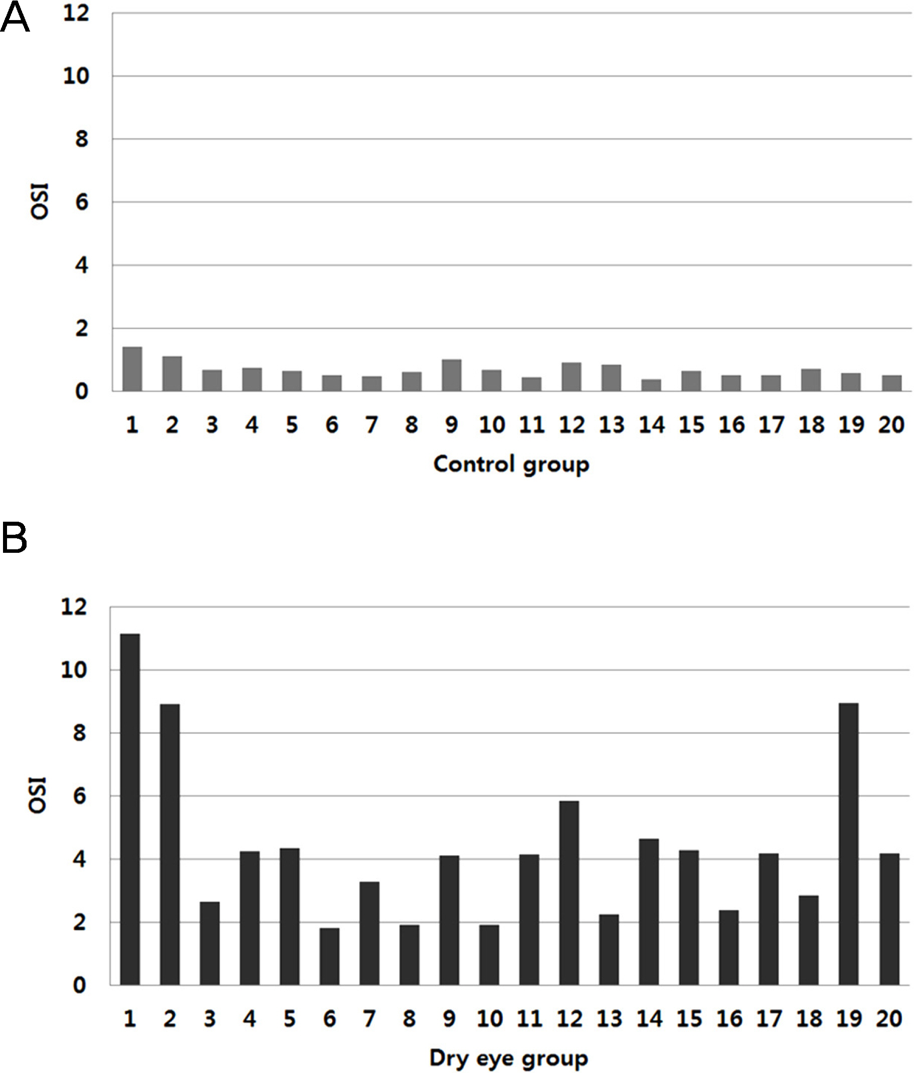
Figure 1. Mean objective scatter index (OSI) value obtained from double-pass image series of all eyes in control group (A) and dry eye group (B).
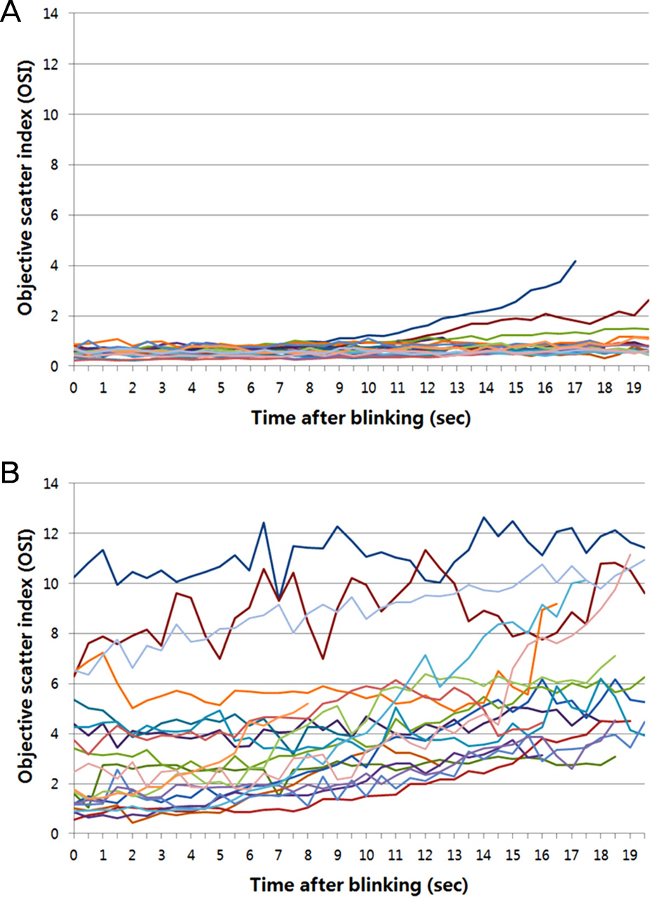
Figure 2. Continuous measured objective scatter index (OSI) in control group (A) and in dry eye group (B). OSI is larger and more variable in dry eye patients.
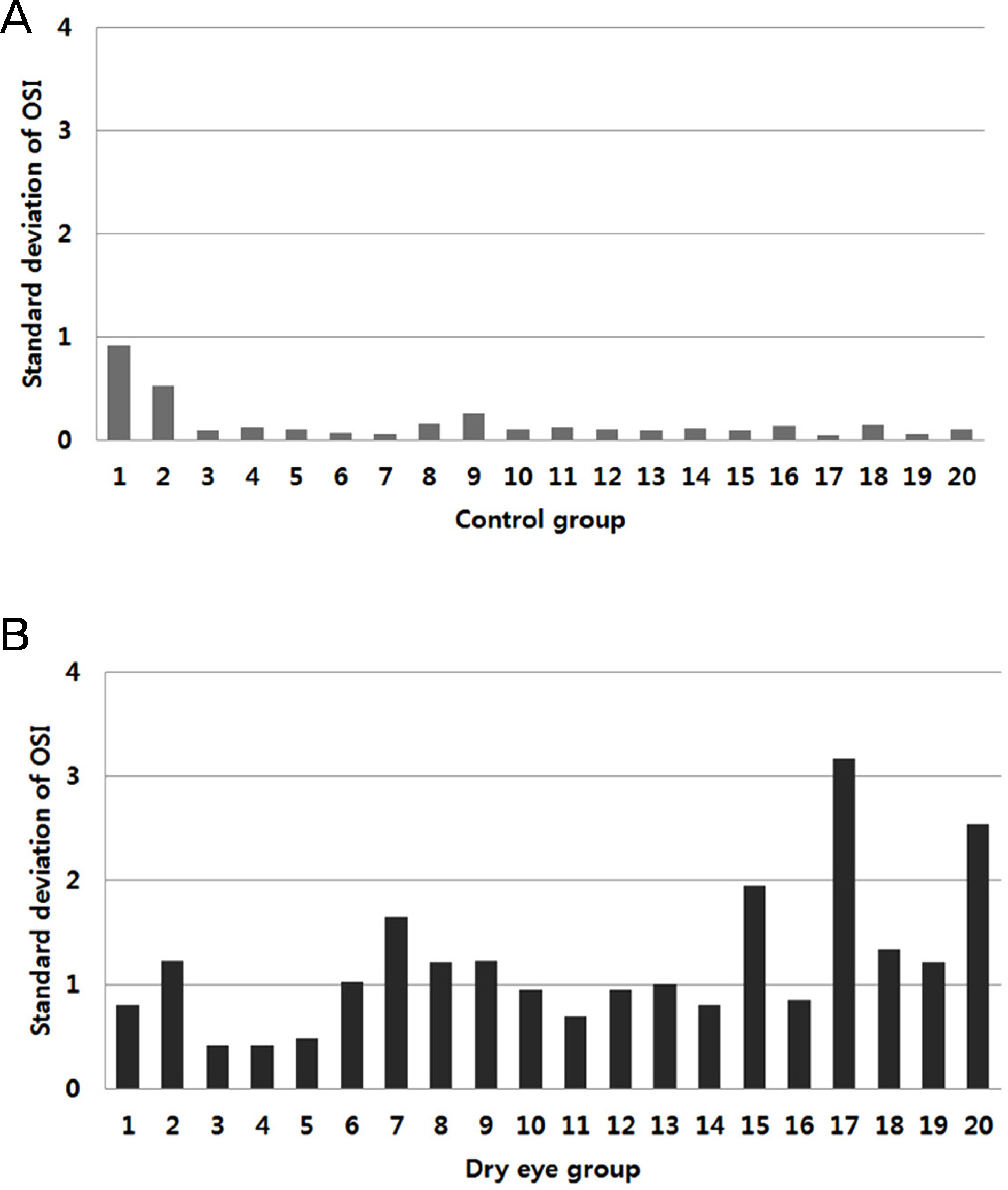
Figure 3. Standard deviation of objective scatter index (OSI) value obtained from double-pass image series of all eyes in control group (A) and dry eye group (B).

Figure 4. Result papers from continuous recording of double pass retinal image. (A) Normal control patient without dry eye symptoms. (B) Mild dry eye syndrome (DES) patient. (C) Severe DES patient with filamentary keratitis. OSI = objective scatter index.
Reference
-
References
1. Montés-Micó R. Role of the tear film in the optical quality of the human eye. J Cataract Refract Surg. 2007; 33:1631–5.
Article2. Tutt R, Bradley A, Begley C, Thibos LN. Optical and visual impact of tear break-up in human eyes. Invest Ophthalmol Vis Sci. 2000; 41:4117–23.3. Thibos LN, Hong X. Clinical applications of the Shack-Hartmann aberrometer. Optom Vis Sci. 1999; 76:817–25.
Article4. The definition and classification of dry eye disease: report of the Definition and Classification Subcommittee of the International Dry Eye WorkShop (2007). Ocul Surf. 2007; 5:75–92.5. Choi SH, Shin YI. Changes in higher order aberration according to tear-film instability analyzed by continuous measurement using wavefront. J Korean Ophthalmol Soc. 2012; 53:1076–80.
Article6. Koh S, Maeda N, Kuroda T, et al. Effect of tear film break-up on higher-order aberrations measured with wavefront sensor. Am J Ophthalmol. 2002; 134:115–7.
Article7. Benito A, Pérez GM, Mirabet S, et al. Objective optical assessment of tear-film quality dynamics in normal and mildly symptomatic dry eyes. J Cataract Refract Surg. 2011; 37:1481–7.
Article8. Schiffman RM, Christianson MD, Jacobsen G, et al. Reliability and validity of the Ocular Surface Disease Index. Arch Ophthalmol. 2000; 118:615–21.
Article9. Methodologies to diagnose and monitor dry eye disease: report of the Diagnostic Methodology Subcommittee of the International Dry Eye WorkShop (2007). Ocul Surf. 2007; 5:108–52.10. Norn MS. Desiccation of the precorneal film. I. Corneal wetting-time. Acta Ophthalmol (Copenh). 1969; 47:865–80.11. Lemp MA. Report of the National Eye Institute/Industry workshop on Clinical Trials in Dry Eyes. CLAO J. 1995; 21:221–32.12. Mengher LS, Bron AJ, Tonge SR, Gilbert DJ. Effect of fluorescein instillation on the pre-corneal tear film stability. Curr Eye Res. 1985; 4:9–12.
Article13. Patel S, Murray D, McKenzie A, et al. Effects of fluorescein on tear breakup time and on tear thinning time. Am J Optom Physiol Opt. 1985; 62:188–90.
Article14. Lee JH, Hyun PM. The reproducibility of the Schirmer test. Korean J Ophthalmol. 1988; 2:5–8.
Article15. Cho P, Yap M. Schirmer test. I. A review. Optom Vis Sci. 1993; 70:152–6.
Article16. Chen JJ, Rao K, Pflugfelder SC. Corneal epithelial opacity in dysfunctional tear syndrome. Am J Ophthalmol. 2009; 148:376–82.
Article17. Choi KW, Moon SW, Joo MJ. Wavefront aberration changes after the instillation of artificial tear in dry eyes. J Korean Ophthalmol Soc. 2006; 47:186–91.18. Saad A, Saab M, Gatinel D. Repeatability of measurements with a double-pass system. J Cataract Refract Surg. 2010; 36:28–33.
Article19. Montés-Micó R, Alió JL, Muñoz G, et al. Postblink changes in total and corneal ocular aberrations. Ophthalmology. 2004; 111:758–67.20. Montés-Micó R, Alió JL, Muñoz G, Charman WN. Temporal changes in optical quality of air-tear film interface at anterior cornea after blink. Invest Ophthalmol Vis Sci. 2004; 45:1752–7.21. The epidemiology of dry eye disease: report of the Epidemiology Subcommittee of the International Dry Eye WorkShop (2007). Ocul Surf. 2007; 5:93–107.22. Diaz-Valle D, Arriola-Villalobos P, García-Vidal SE, et al. Effect of lubricating eyedrops on ocular light scattering as a measure of vision quality in patients with dry eye. J Cataract Refract Surg. 2012; 38:1192–7.
Article
- Full Text Links
-
- Actions
-
Cited
- CITED
-
- Close
- Share
-
- Similar articles
-
- Points to consider when developing drugs for dry eye syndrome
- The significance of tear film break-up time in the diagnosis of dry eye syndrome
- Bacterial Conjuntival Flora in Dry Eye Patients
- Clinical Usefulness of a Thermal-Massaging System for Treatment of Dry Eye with Meibomian Gland Dysfunction
- Dry Eye Assessment of Patients Undergoing Endoscopic Dacryocystorhinostomy for Nasolacrimal Duct Obstruction Combined with Dry Eye Syndrome
