J Korean Ophthalmol Soc.
2013 Jan;54(1):33-37. 10.3341/jkos.2013.54.1.33.
Corneal Endothelial Changes after Laser-Assisted Subepithelial Keratomileusis
- Affiliations
-
- 1Department of Ophthalmology and Visual Science, The Catholic University of Korea College of Medicine, Seoul, Korea. eyedoc@catholic.ac.kr
- KMID: 2216446
- DOI: http://doi.org/10.3341/jkos.2013.54.1.33
Abstract
- PURPOSE
In order to investigate the safety of laser-assisted subepithelial keratomileusis (LASEK), corneal endothelial cells before and after the LASEK procedure were evaluated.
METHODS
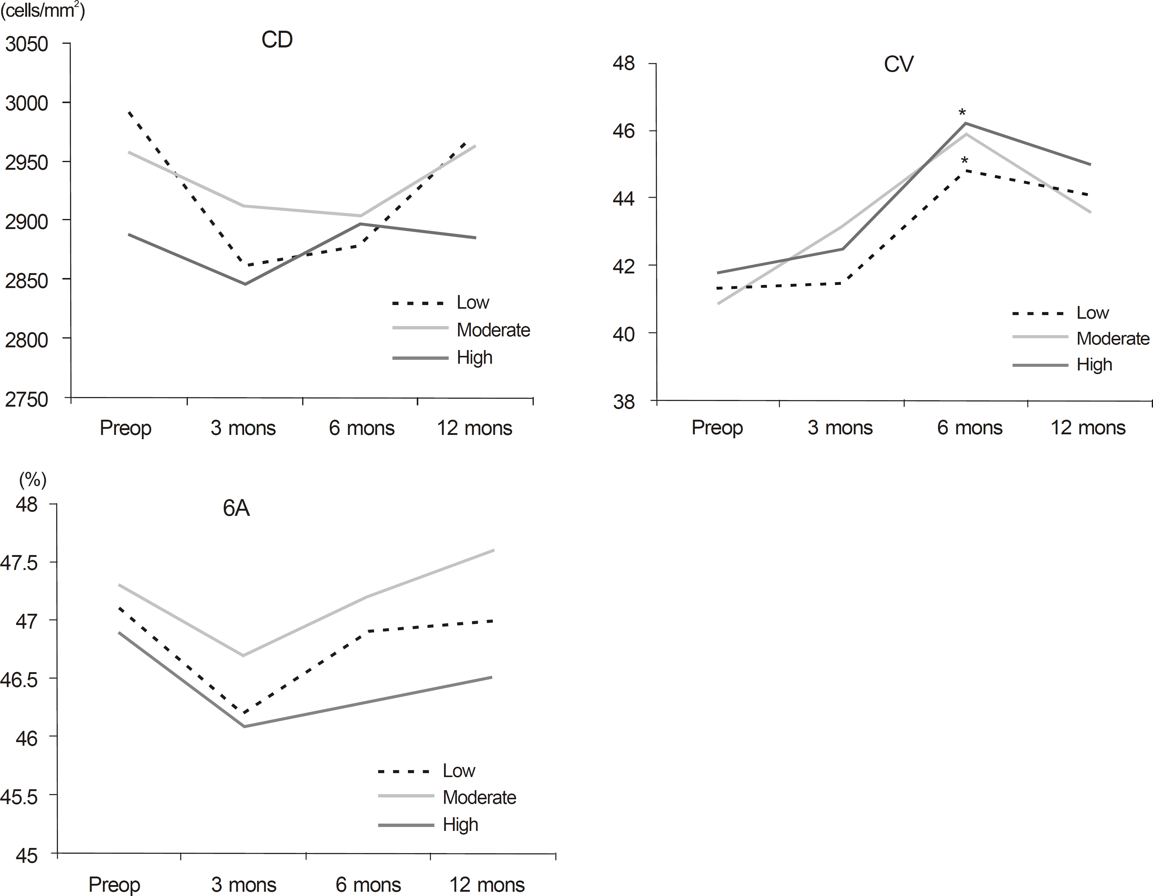
Thirty-six patients (72 eyes) who underwent LASEK between June 2010 and May 2011 were included in the present study. Parameters included corneal endothelial cell density (CD), coefficient of variation of the cell area (CV), and percentage of hexagonal cells (6A) which were all obtained by a specular microscope (Noncon ROBO sp 8000, Konan, Japan) before and 3, 6, and 12 months after LASEK.
RESULTS
Preoperative CD was 2952 +/- 352 cells/mm2, and postoperative CD did not significantly change at 3, 6, and 12 months. Preoperative CV and 6A and postoperative CV and 6A at 12 months were not significantly different. Furthermore, correlation between change in corneal endothelial cell and degree of myopia correction was not statistically significant.
CONCLUSIONS
LASEK appears to be a safe procedure for corneal endothelial cells over an extended period.
Keyword
MeSH Terms
Figure
Cited by 1 articles
-
Corneal Endothelial Cell Changes after LASEK and M-LASEK
Seung Jae Lee, Damho Lee, Haksu Kyung
J Korean Ophthalmol Soc. 2013;54(10):1501-1507. doi: 10.3341/jkos.2013.54.10.1501.
Reference
-
References
1. Camellin M. Laser epithelial keratomileusis for myopia. J Refract Surg. 2003; 19:666–70.
Article2. Camellin M. M Cimberle. LASEK may offer the advantages of both LASIK and PRK. Ocular Surgery News. 1999; 28.3. Teus MA, de Benito-Llopis L, Sánchez-Pina JM. LASEK versus LASIK for the correction of moderate myopia. Optom Vis Sci. 2007; 84:605–10.
Article4. Kim JK, Kim SS, Lee HK, et al. Laser in situ keratomileusis versus laser-assisted subepithelial keratectomy for the correction of high myopia. J Cataract Refract Surg. 2004; 30:1405–11.
Article5. Autrata R, Rehurek J. Laser-assisted subepithelial keratectomy and photorefractive keratectomy for the correction of hyperopia. Results of a 2-year follow-up. J Cataract Refract Surg. 2003; 29:2105–14.
Article6. Kim HJ, Joo CK. Clinical results of laser epithelial keratomileusis and laser in situ keratomileusis for morderate and high myopia. J Korean Ophthalmol Soc. 2003; 44:1159–64.7. Kymionis GD, Tsiklis NS, Astyrakakis N, et al. Eleven-year fol-low-up of laser in situ keratomileusis. J Cataract Refract Surg. 2007; 33:191–6.
Article8. Trokel SL, Srinivasan R, Braren B. Excimer laser surgery of the cornea. Am J Ophthalmol. 1983; 96:710–5.
Article9. Smith RT, Waring GO 4th, Durrie DS, et al. Corneal endothelial cell density after femtosecond thin-flap LASIK and PRK for abdominal: a contralateral eye study. J Refract Surg. 2009; 25:1098–102.10. Tsiklis NS, Kymionis GD, Pallikaris AI, et al. Endothelial cell abdominal after photorefractive keratectomy for moderate myopia using a 213 nm solid-state laser system. J Cataract Refract Surg. 2007; 33:1866–70.11. Collins MJ, Carr JD, Stulting RD, et al. Effects of laser in situ abdominal (LASIK) on the corneal endothelium 3 years postoperatively. Am J Ophthalmol. 2001; 131:1–6.12. Kim TG, Joo CK. abdominal changes of corneal endothelium after LASIK. J Korean Ophthalmol Soc. 1999; 40:46–54.13. Waltman SR, Yarian D, Hart W Jr, Becker B. Corneal endothelial changes with long-term topical epinephrine therapy. Arch Ophthalmol. 1977; 95:1357–8.
Article14. Perkins TW, Kumar A, Kiland JA. Corneal decompensation abdominal bleb revision with absolute alcohol: clinical pathological correlation. Arch Ophthalmol. 2006; 124:738–41.15. Zhou J, Lu S, Dai J, et al. abdominal corneal endothelial changes after laser-assisted subepithelial keratectomy. J Int Med Res. 2010; 38:1484–90.16. O'Connor J, O'Keeffe M, Condon PI. Twelve-year follow-up of photorefractive keratectomy for low to moderate myopia. J Refract Surg. 2006; 22:871–7.17. Marshall J, Trokel S, Rothery S, Schubert H. An ultrastructural study of corneal incisions induced by an excimer laser at 193 nm. Ophthalmology. 1985; 92:749–58.
Article18. Gaster RN, Binder PS, Coalwell K, et al. Corneal surface ablation by 193 nm excimer laser and wound healing in rabbits. Invest Ophthalmol Vis Sci. 1989; 30:90–8.19. Hanna KD, Pouliquen Y, Waring GO 3rd, et al. Corneal stromal wound healing in rabbits after 193-nm excimer laser surface ablation. Arch Ophthalmol. 1989; 107:895–901.
Article20. Cennamo G, Rosa N, Guida E, et al. Evaluation of corneal abdominal and endothelial cells before and after excimer laser photo-refractive keratectomy. J Refract Corneal Surg. 1994; 10:137–41.21. Pérez-Santonja JJ, Sakla HF, Gobbi F, Alió JL. Corneal endothelial changes after laser in situ keratomileusis. J Cataract Refract Surg. 1997; 23:177–83.
Article22. Nishida T, Saika S. Cornea and sclera: anatomy and physiology. Krachmer JH, Mannis MJ, Holland EJ, editors. Cornea. 3rd ed.St. Louis: MO Mosby;2011. p. 15–6.23. Patel SV, Bourne WM. Corneal endothelial cell loss 9 years after excimer laser keratorefractive surgery. Arch Ophthalmol. 2009; 127:1423–7.
Article24. Pallikaris IG, Siganos DS. Laser in situ keratomileusis to treat abdominal: early experience. J Cataract Refract Surg. 1997; 23:39–49.25. Han HS, Jung HR, Kim HM. Corneal endothelial changes after abdominal assisted in situ keratomileusis. J Korean Ophthalmol Soc. 1997; 38:1510–6.
- Full Text Links
- Actions
-
Cited
- CITED
-
- Close
- Share
- Similar articles
-
- Corneal Endothelial Changes after Laser Assisted In Situ Keratomileusis
- Long-term Outcome of Combined Laser-assisted Subepithelial Keratomileusis and Accelerated Corneal Crosslinking for Myopia
- Corneal Endothelial Damage after Deep Excimer Laser Ablation
- Surgical treatment of presbyopia
- Short-Term Changes of Corneal Endothelium after LASIK


