J Korean Ophthalmol Soc.
2015 Feb;56(2):300-303. 10.3341/jkos.2015.56.2.300.
A Case of Cyclodialysis Cleft with Hypotony during Ahmed Valve Implantation Surgery
- Affiliations
-
- 1Department of Ophthalmology, Catholic University of Daegu School of Medicine, Daegu, Korea. jwkim@cu.ac.kr
- KMID: 2216018
- DOI: http://doi.org/10.3341/jkos.2015.56.2.300
Abstract
- PURPOSE
To report a case of cyclodialysis cleft with hypotony during Ahmed valve implantation.
CASE SUMMARY
A 47-year-old male was referred for uncontrolled intraocular pressure (IOP) in the right eye. The patient had a history of ocular trauma and traumatic hyphema. He underwent pars plana vitrectomy, phacoemulsification and intraocular lens implantation 1 month prior due to rhegmatogenous retinal detachment. At the end of the Ahmed valve implantation surgery, the eye was hypotonic despite a deep anterior chamber. The hypotony continued and choroidal effusion developed. Anterior segment optical coherence tomography and gonioscopic examinations revealed small cyclodialysis clefts. After medical treatment with cycloplegics and steroids, choroidal effusion disappeared and IOP was normalized.
CONCLUSIONS
Patients with previous ocular trauma or surgery could be prone to developing cyclodialysis cleft with hypotony. Inadvertent cyclodialysis cleft with hypotony can be treated with cycloplegics and steroids.
Keyword
MeSH Terms
Figure
-
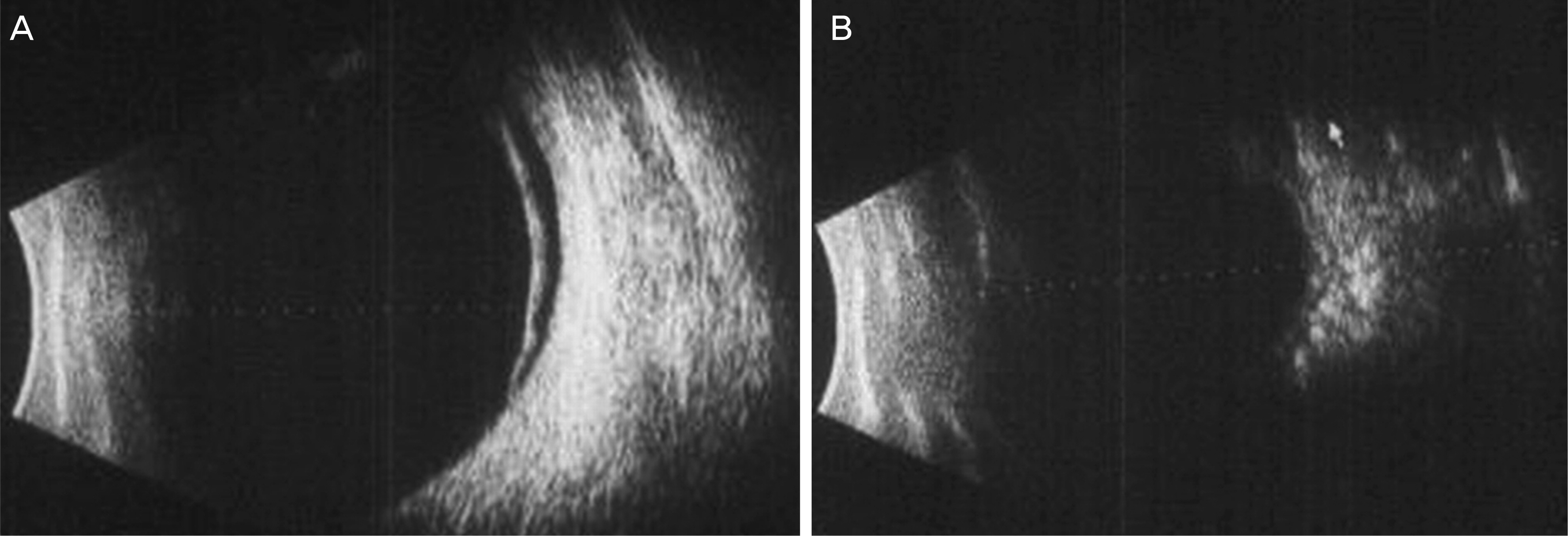
Figure 1. B-scan ultrasonography reveals shallow choroidal detachment (A) and effusion (B).
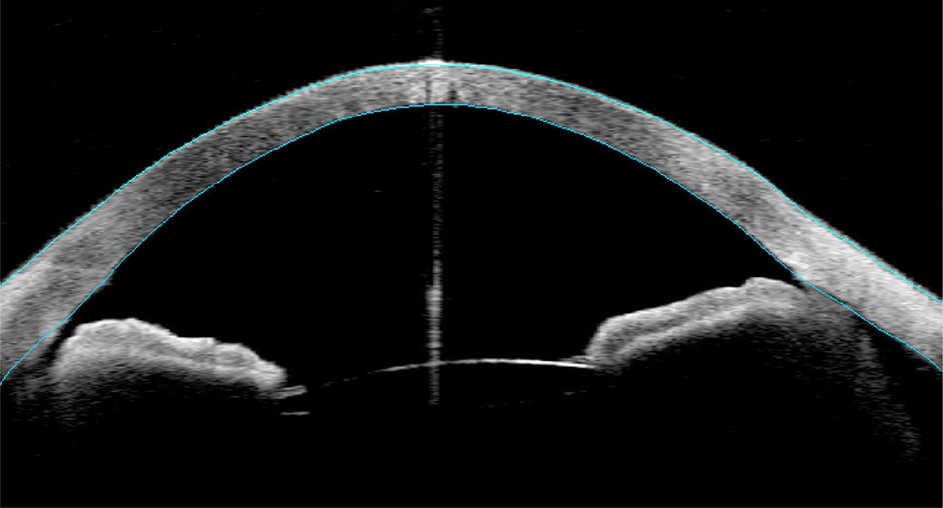
Figure 2. Anterior segment OCT reveals cyclodialysis cleft (left). OCT = optical coherence tomography.
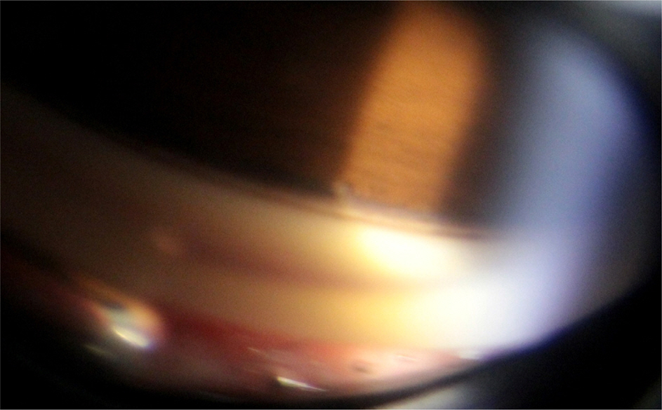
Figure 3. Gonioscopy reveals suspected small cyclodialysis at the corresponding region of anterior segment OCT. OCT = optical coherence tomography.
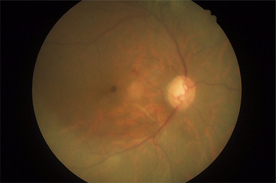
Figure 4. Choroidal detachment disappeared after medical treatment for 2 weeks.
Reference
-
References
1. Fuchs E. Ablösung der Aderhaut nach Staaroperation. Graefes Arch Clin Exp Ophthalmol. 1900; 51:199–224.
Article2. Heine L. Die Cyklodialyse, eine neue glaucomoperation. Deutsche Med Wehnschr. 1905; 31:824–6.3. Maffett MJ, O'Day DM. Cyclodialysis cleft following a scleral tunnel incision. Ophthalmic Surg. 1994; 25:387–8.
Article4. Small EA, Solomon JM, Prince AM. Hypotonus cyclodialysis cleft following suture fixation of a posterior chamber intraocular lens. Ophthalmic Surg. 1994; 25:107–9.
Article5. Parnes RE, Dailey JR, Aminlari A. Hypotonus cyclodialysis cleft following anterior chamber intraocular lens removal. Ophthalmic Surg. 1994; 25:386–7.
Article6. Espana EM, Tello C, Liebmann JM, Ritch R. Cyclodialysis cleft secondary to removal of an anterior chamber phakic intraocular lens. J Cataract Refract Surg. 2007; 33:542–4.
Article7. Shah VA, Majji AB. Ultrasound biomicroscopic documentation of traumatic cyclodialysis cleft closure with hypotony by medical therapy. Eye (Lond). 2004; 18:857–8.
Article8. Aminlari A, Callahan CE. Medical, Laser, and Surgical Management of Inadvertent CyclodialysisCleft With Hypotony. Arch Ophthalmol. 2004; 122:396–8.
Article9. Bauer B. Argon laser photocoagulation of cyclodialysis clefts after cataract surgery. Acta Ophthalmol Scand. 1995; 73:283–4.
Article10. Harbin TS Jr. Treatment of cyclodialysis clefts with argon laser photocoagulation. Ophthalmology. 1982; 89:1082–3.
Article11. Alward WL, Hodapp EA, Parel JM, Anderson DR. Argon laser en-dophotocoagulator closure of cyclodialysis clefts. Am J Ophthalmol. 1988; 106:748–9.
Article12. Hwang JM, Ahn K, Kim C. . Ultrasonic biomicroscopic evaluation of cyclodialysis before and after direct cyclopexy. Arch Ophthalmol. 2008; 126:1222–5.
Article13. Mandava N, Kahook MY, Mackenzie DL, Olson JL. Anterior scleral buckling procedure for cyclodialysis cleft with chronic hypotony. Ophthalmic Surg Lasers Imaging. 2006; 37:151–3.
Article14. Yuen NS, Hui SP, Woo DC. New method of surgical repair for 360-degree cyclodialysis. J Cataract Refract Surg. 2006; 32:13–7.
Article15. Hoerauf H, Roider J, Laqua H. Treatment of traumatic cyclodialysis with vitrectomy, cryotherapy, and gas endotamponade. J Cataract Refract Surg. 1999; 25:1299–301.
Article16. Takaya K, Suzuki Y, Nakazawa M. Four cases of hypotony maculopathy caused by traumatic cyclodialysis and treated by vi-trectomy, cryotherapy, and gas tamponade. Graefes Arch Clin Exp Ophthalmol. 2006; 244:855–8.
Article17. Kim KH, Song BJ, Choi YI. Hypotony with cyclodialysis after blunt trauma to the eye. J Korean Ophthalmol Soc. 1997; 38:121–8.18. Moon HS, Nam DH, Kim SW. Treatment of hypotony retinopathy with cyclodialysis cleft by intravitreal gas injection. J Korean Ophthalmol Soc. 2006; 47:319–22.
- Full Text Links
-
- Actions
-
Cited
- CITED
-
- Close
- Share
-
- Similar articles
-
- A Case of Cyclodialysis Cleft Treated with Argon Laser Photocoagulation
- Hypotony with Cyclodialysis after Blunt Trauma to the Eye
- Clinical Features and Histopathological Findings of Traumatic Cyclodialysis Clefts
- A Case of Macular Serous Retinal Detachment after Ahmed Valve Implantation in an Eye with Pachychoroid
- A Case of Traumatic Cyclodialysis Cleft Diagnosed by Ultrasound Biomicroscopy
