Clinical Characteristics of Acute Zonal Occult Outer Retinopathy
- Affiliations
-
- 1Department of Ophthalmology, Seoul National University Bundang Hospital, Seoul National University College of Medicine, Seongnam, Korea. hjm@snu.ac.kr
- KMID: 2213242
- DOI: http://doi.org/10.3341/jkos.2016.57.3.413
Abstract
- PURPOSE
To investigate the clinical characteristics of patients with acute zonal occult outer retinopathy (AZOOR).
METHODS
Medical records of 13 patients who visited Seoul National University Bundang Hospital from May, 2003 to May, 2015 and diagnosed with AZOOR were retrospectively reviewed.
RESULTS
Thirteen patients (11 women and 2 men), with a mean age of 28.5 ± 11.4 years were followed for 42.8 ± 30.3 months. Visual field loss, photopsia, and blurred vision were common complaints. Initially, four patients had bilateral disease and seven patients showed bilateral involvement at the last visit. Mean best corrected visual acuity of involved eyes (BCVA) was 0.75 ± 0.32 (log MAR). Among 20 eyes with AZOOR, BCVA was 20/40 or better in 17 eyes (85.0%). The mean spherical equivalent was -4.59 ± 3.23 diopters (D), and 15 eyes (75.0%) had myopia less than -2.00 D. Nine eyes of seven patients (34.6%) had abnormal fundus findings. All patients underwent full field electroretinogram (ERG) or multifocal ERG and a visual field test. Thirteen patients (100.0%) showed a decreased response in ERG and visual field defects presented in every patient. With respect to the visual field test, 10 eyes (50.0%) showed improvement, 6 eyes (30.0%) had stationary status, and the progression of the visual field defect was observed in 4 eyes (20.0%). Among 13 patients, 4 (30.8%) patients showed flu-like symptom, 4 (30.8%) patients had fatigue, 2 (15.4%) patients had nausea, and 2 (15.4%) patients showed headache.
CONCLUSIONS
AZOOR should be considered as one of the differential diagnoses, especially in female patients with myopia who show photopsia or visual field defects. ERG and visual field tests are necessary to confirm a decrease in retinal function and visual field loss. Central vision is preserved in most cases and recovery of visual field defect occurs often.
MeSH Terms
Figure
-

Figure 1. Fundus photography and spectral-domain optical coherence tomography examination. Fundus photograph (A) of the left eye shows normal posterior pole. Corresponding image of spectral-domain optical coherence tomography (B) shows disruption (arrowheads) of the internal segment-outer segment junction and cone outer segment tip line at temporal peripapillary area.
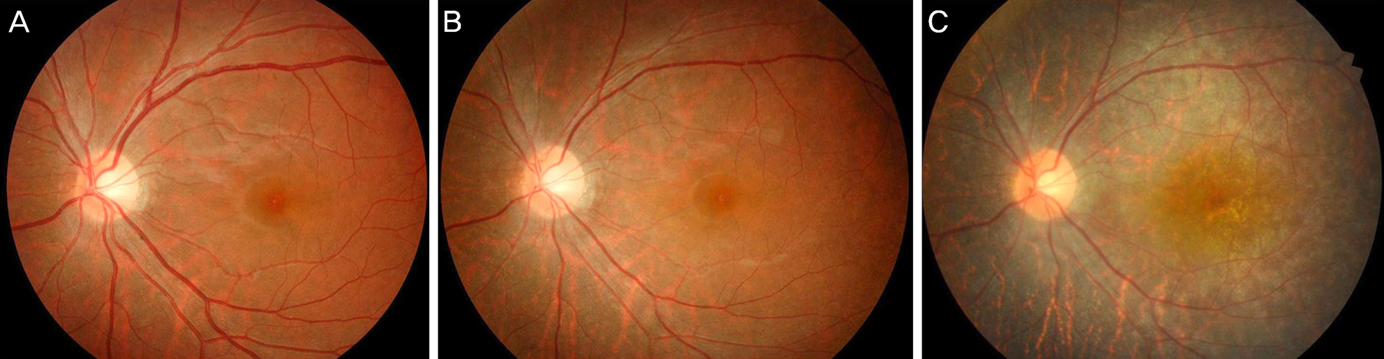
Figure 2. Fundus photography showing retinal degeneration. Fundus photograph of the left eye shows normal appearance one month after presentation (A). Seven months later after then, there is mild hypopigmentation of the retinal pigmented epithelium (RPE) around peripapillary area (B). At 69 months from the onset, it is observed that generalized severe RPE atrophy and depigmentation (C).

Figure 3. Visual field test and electroretinogram. Goldmann perimetry (target I4) shows central scotoma of the left eye (A). Decreased amplitudes (circles with dotted line) corresponding visual field defect is observed in multifocal electroretinogram (ERG) (B). Standard full-field ERG reveals slightly decreased rod and maximal combined response in left eye compared to those of the right eye (C). Rt = right; Lt = left; Diff = difference.
Reference
-
References
1. Gass JD. Acute zonal occult outer retinopathy. Donders Lecture: The Netherlands Ophthalmological Society, Maastricht, Holland, June 19, 1992. J Clin Neuroophthalmol. 1993; 13:79–97.2. Gass JD, Agarwal A, Scott IU. Acute zonal occult outer retinopathy: a long-term follow-up study. Am J Ophthalmol. 2002; 134:329–39.
Article3. Choi HJ, Hwang JM. A case report of acute zonal occult outer retinopathy. J Korean Ophthalmol Soc. 2003; 44:1384–91.4. Kim JH, Kim MJ, Park HS. A case report of acute zonal occult outer retinopathy showing acute idiopathic blind spot enlargement. J Korean Ophthalmol Soc. 2011; 52:364–72.
Article5. You JY, Chung H, Kim HC. Acute zonal occult outer retinopathy, responsive to an immunosuppressive agent: a case report. J Korean Ophthalmol Soc. 2011; 52:492–501.
Article6. Sperduto RD, Seigel D, Roberts J, Rowland M. Prevalence of myopia in the United States. Arch Ophthalmol. 1983; 101:405–7.
Article7. Watzke RC, Packer AJ, Folk JC, et al. Punctate inner choroidopathy. Am J Ophthalmol. 1984; 98:572–84.
Article8. Kim EC, Morgan IG, Kakizaki H, et al. Prevalence and risk factors for refractive errors: Korean National Health and Nutrition Examination Survey 2008-2011. PLoS One. 2013; 8:e80361.
Article9. Yoo YC, Kim JM, Park KH, et al. Refractive errors in a rural Korean adult population: the Namil Study. Eye (Lond). 2013; 27:1368–75.
Article10. Fujiwara T, Imamura Y, Giovinazzo VJ, Spaide RF. Fundus autofluorescence and optical coherence tomographic findings in acute zonal occult outer retinopathy. Retina. 2010; 30:1206–16.
Article11. Mkrtchyan M, Lujan BJ, Merino D, et al. Outer retinal structure in patients with acute zonal occult outer retinopathy. Am J Ophthalmol. 2012; 153:757–68. 768.e1.
Article12. Park KH, Woo SJ, Lee JJ, Hwang JM. Multifocal electroretinogram changes and normal Ganzfeld-ERG in patients with AZOOR. Neuro-Ophthalmology. 2008; 32:291–4.
Article13. Arai M, Nao-i N, Sawada A, Hayashida T. Multifocal electroretinogram indicates visual field loss in acute zonal occult outer retinopathy. Am J Ophthalmol. 1998; 126:466–9.
Article14. Fekrat S, Wilkinson CP, Chang B, et al. Acute annular outer retinopathy: report of four cases. Am J Ophthalmol. 2000; 130:636–44.
Article15. Song ZM, Sheng YJ, Chen QS, et al. Clinical characteristics of acute zonal occult outer retinopathy in Chinese patients. Ophthalmologica. 2008; 222:149–56.
Article16. Tang J, Stevens RA, Okada AA, et al. Association of antiretinal antibodies in acute annular outer retinopathy. Arch Ophthalmol. 2008; 126:130–2.
Article17. Spaide RF, Koizumi H, Freund KB. Photoreceptor outer segment abnormalities as a cause of blind spot enlargement in acute zonal occult outer retinopathy-complex diseases. Am J Ophthalmol. 2008; 146:111–20.
Article18. Yasuda K, Shimura M, Noro M, et al. Clinical course of acute retinal zonal occult outer retinopathy in visual field and multifocal electroretinogram. Br J Ophthalmol. 1999; 83:1089–90.
- Full Text Links
-
- Actions
-
Cited
- CITED
-
- Close
- Share
-
- Similar articles
-
- Acute Zonal Occult Outer Retinopathy Mimicking Retrobulbar Optic Neuritis
- Fulminant Progressive Atypical Acute Zonal Occult Outer Retinitis
- A Case Report of Acute Zonal Occult Outer Retinopathy
- A Case Report of Acute Zonal Occult Outer Retinopathy Showing Acute Idiopathic Blind Spot Enlargement
- Acute Zonal Occult Outer Retinopathy, Responsive to an Immunosuppressive Agent: A Case Report
