Blepharoptosis Repair by Small Cutaneous Incision and Minimal Dissection Technique
- Affiliations
-
- 1Department of Ophthalmology, College of Medicine, The Catholic University of Korea, Seoul, Korea. laty@catholic.ac.kr
- KMID: 2212527
- DOI: http://doi.org/10.3341/jkos.2009.50.8.1146
Abstract
- PURPOSE
To present a simple method of acquired ptosis correction by small-incision minimal dissection technique and assess the results of the operation. METHODS: The charts of 23 patients (29 eyes) with acquired ptosis who underwent ptosis correction by small-incision minimal dissection technique were reviewed. Pre and postoperative MRD1, success rate, complications and reoperation rates were investigated. RESULTS: The average of pre- and postoperative MRD1 were 0.9+/-0.9 mm and 2.7+/-0.8 mm respectively. Of the 17 patients who underwent unilateral surgery, 15 eyes (88.2%) showed successful outcomes, and of the 12 eyes who underwent bilateral surgery, 8 eyes (66.6%), 2 eyes (16.7%), and 2 eyes (16.7%) showed excellent, good, and poor outcomes, respectively. Out of 29 eyes, 25 eyes (86.2%) showed satisfactory results. Two eyelids of unsatisfactory contour were corrected by reoperation. CONCLUSIONS: Although the small-incision minimal dissection technique for ptosis correction is applicable to a restricted group of patients compared to the conventional method, this technique is very useful and efficient, and has many advantages including less tissue damage, bleeding, edema, a short operation time and rapid recovery.
Keyword
Figure
-
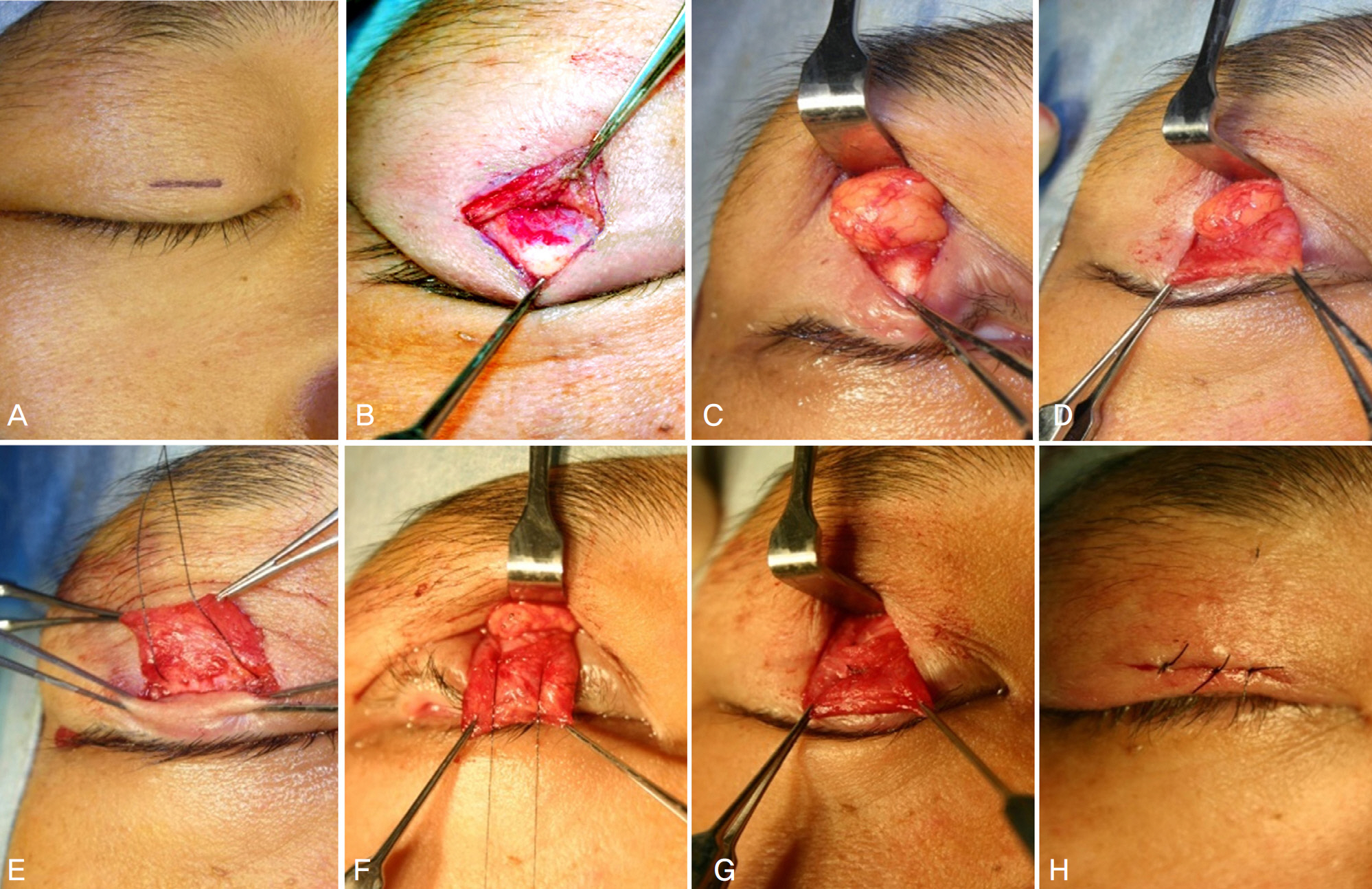
Figure 1. Procedures of small cutaneous incision and minimal dissection technique for ptosis correction. (A) A length of 8 to 10-mm skin marking was drawn along the lid crease above the center of the pupil. (B) Tarsal plate was exposed after dissecting the pretarsal orbicularis. (C) Preaponeurotic fat pad was identified after incising the orbital septum over the tarsal plate. (D) Dissection of levator aponeurosis was carried out to a point at the level of musculo-aponeurotic junction. (E) A partial thickness suture of tarsal plate was placed in a horizontal fashion with more than 5-mm width. (F) The double armed, non absorbable suture was passed through the levator aponeurosis in the appropriate position. (G) The fixation suture was tied after inspecting the eyelid position and contour. (H) Finally, the skin was closed.
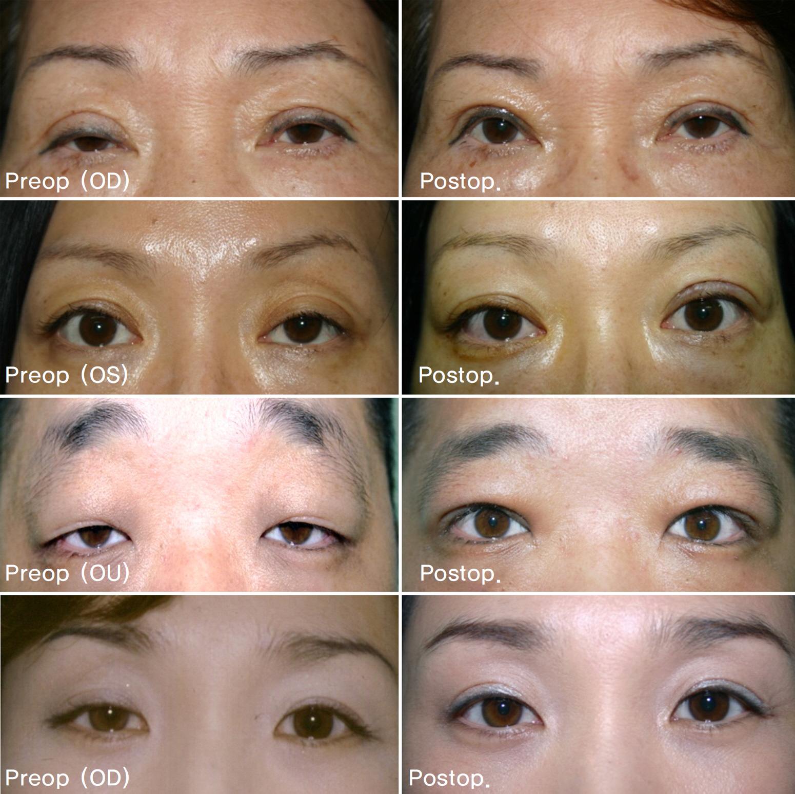
Figure 2. Preoperative and postoperative photographs of patients.
Reference
-
References
1. Chung WS. Treatment of ptosis. Lee SY, Kim YD, Khwarg SI, Kim SJ, editors. Ophthalic plastic and reconstructive surgery. Seoul: NWHS;2004. 1:chap. 6.2. Dresner SC. Ptosis management: A practical approach. WP C, editor. Oculoplastic surgery: The essentials. New York: Thieme Medical Publishers;2001. 1:chap. 6.3. Iliff J, Pacheoco E. Ptosis surgery. Tasman W, Jaeger E, editors. Duane's clinical ophthalmology. Philadelpia: Williams and Wilkins;2001. 5:chap. 72.4. Lucarelli MJ, Lemke BN. Small incision external levator repair: technique and early results. Am J Ophthalmol. 1999; 127:637–44.
Article5. Baroody M, Holds JB, Sakamoto DK, et al. Small incision trans-cutaneous levator aponeurotic repair for blepharoptosis. Ann Plast Surg. 2004; 52:558–61.
Article6. Bernardini FP, de Conciliis C, Devoto MH. Mini-invasive ptosis surgery. Orbit. 2006; 25:111–5.
Article7. Frueh BR, Musch DC, McDonald H. Efficacy and efficiency of a new involutional ptosis correction procedure compared to a traditional aponeurotic approach. Trans Am Ophthalmol Soc. 2004; 102:199–206.8. Frueh BR, Musch DC, McDonald HM. Efficacy and efficiency of a small incision, minimal dissection procedure versus a traditional approach for correcting aponeurotic ptosis. Ophthalmology. 2004; 111:2158–63.9. Lee TS, Nam DH. Levator function, modified Fasanella-Servat operation, ptosis degree, srugical results. J Korean Ophthalmol Soc. 1999; 40:248–52.10. Moon HS, Lee JH, Baek SH. The frontalis sling operation using preserved fascia lata: modified crawford technique. J Korean Ophthalmol Soc. 2005; 46:10–5.11. Carter SR, Meecham WJ, Seiff SR. Silicone frontalis slings for the correction of blepharoptosis: indications and efficacy. Ophthalmology. 1996; 103:623–30.12. Everbusch O. Zur Operation der congenitalen Blepharoptosis. Klin Monatsbl Augenheilkd. 1883; 21:100–7.13. Jones LT, Quickert MH, Wobig JL. The cure of ptosis by aponeurotic repair. Arch Ophthalmol. 1975; 93:629–34.
Article14. Liu D. Ptosisrepair by single suture aponeurotic tuck. Surgical technique and long-term results. Ophthalmology. 1993; 100:251–9.15. Meltzer MA, Elahi E, Taupeka P, Flores E. A simplified technique of ptosis repair using a single adjustable suture. Ophthalmology. 2001; 108:1889–92.
Article16. Jeong S. Clinical study of simple levator resection in ptosis patients. J Korean Ophthalmol Soc. 2002; 43:551–5.17. Baek SH, Park MS, Park HJ. The effect of simplified single suture aponeurotic tuck in ptosis patients. J Korean Ophthalmol Soc. 2003; 44:1011–6.18. Lee JW, Shin DS, Lee KW. Small incision for ptosis repair. J Korean Ophthalmol Soc. 2008; 49:721–6.
Article
- Full Text Links
-
- Actions
-
Cited
- CITED
-
- Close
- Share
-
- Similar articles
-
- Blepharoptosis Repair through the Small Orbital Septum Incision and Minimal Dissection Technique in Patients with Coexisting Dermatochalasis
- Cases of Mild Ptosis Correction with Suture-Method
- Minimal-incision Tenorrhaphy in Flexor Tendon Injury
- Small Incision for Ptosis Repair
- A Comparative Study of Surgical Treatment in the Ruptured Achilles Tendon: Minimal incision and Open repair
