J Korean Ophthalmol Soc.
2009 Jul;50(7):1066-1070. 10.3341/jkos.2009.50.7.1066.
Sclerotomy-related Retinal Breaks in Vitrectomy for Proliferative Diabetic Retinopathy: 20- vs 23-Gauge Systems
- Affiliations
-
- 1Department of Ophthalmology, Gachon University Gil Medical Center, Incheon, Korea. eyedawns@gilhospital.com
- KMID: 2212495
- DOI: http://doi.org/10.3341/jkos.2009.50.7.1066
Abstract
- PURPOSE
To compare the rate of intraoperative sclerotomy-related retinal breaks (SRRB) between 20- and 23-gauge vitrectomy for proliferative diabetic retinopathy (PDR). METHODS: Medical records of 62 consecutive eyes of 54 patients who underwent 20-gauge pars plana vitrectomy (PPV) and 63 consecutive eyes of 55 patients who received 23-gauge transconjunctival sutureless vitrectomy were retrospectively reviewed. RESULTS: Three (4.8%) out of 62 eyes in the 20-gauge group had SRRB and 1 (1.6%) eye developed retinal detachment at 4 months postoperatively, while 2 (3.2%) out of 63 eyes in the 23-gauge group had SRRB and 1 (1.6%) eye developed retinal detachment at 3 months postoperatively. CONCLUSIONS: There were no significant differences in the rates of sclerotomy-related retinal breaks and sclerotomy-related retinal detachments between 20-gauge PPV and 23-gauge PPV for PDR.
Keyword
MeSH Terms
Figure
-

Figure 1. 23-gauge microcannula (DORC, Holland). The length (without its head) is 4 mm, the internal diameter is 0.65 mm, and the external diameter is 0.75 mm. The external opening is funnel shaped.
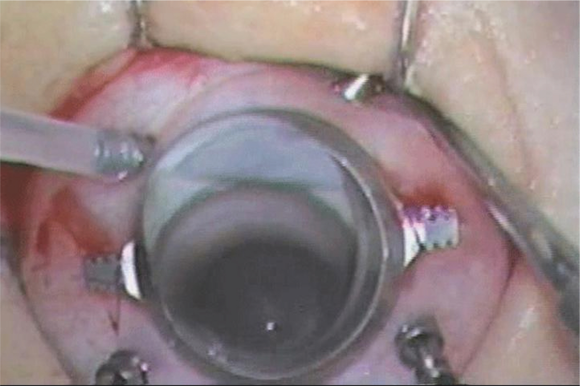
Figure 2. Intraoperative photograph of the procedure searching sclerotomy-related retinal breaks by 360° scleral depression.
Cited by 1 articles
-
Effect of 23-gauge Sutureless Vitrectomy & Preoperative Bevacizumab on Results of Diabetic Vitrectomy
Dae Heon Han, Hee Jin Sohn, Dae Young Lee, Dong Heun Nam
J Korean Ophthalmol Soc. 2011;52(3):285-292. doi: 10.3341/jkos.2011.52.3.285.
Reference
-
References
1. Carter JB, Michels RG, Glaser BM, De Bustros S. Iatrogenic retinal breaks complicating pars plana vitrectomy. Ophthalmology. 1990; 97:848–53.
Article2. Sjaarda RN, Glaser BM, Thompson JT, et al. Distribution of iatrogenic retinal breaks in macular hole surgery. Ophthalmology. 1995; 102:1387–92.
Article3. Al-Harthi E, Abboud EB, Al-Dhibi H, Dhindsa H. Incidence of sclerotomy-related retinal breaks. Retina. 2005; 25:281–4.
Article4. Tognetto D, di Lauro MT, Fanni D, et al. Iatrogenic retinal traumas in ophthalmic surgery. Graefes Arch Clin Exp Ophthalmol. 2008; 246:1361–72.
Article5. Singh DV, Pal N, Azad RV. Incidence of early and late sclerotomy-related retinal breaks in patients who underwent pars plana vitrectomy. Retina. 2006; 26:251.6. Scartozzi R, Bessa AS, Gupta OP, Regillo CD. Intraoperative sclerotomy-related retinal breaks for macular surgery, 20-vs 25-gauge vitrectomy systems. Am J Ophthalmol. 2007; 143:155–6.7. Sabti K, Kapusta M, Mansour M, et al. Ultrasound biomicroscopy of sclerotomy sites: the effect of vitreous shaving around sclerotomy sites during pars plana vitrectomy. Retina. 2001; 21:464–8.8. Territo C, Gieser JP, Wilson CA, Anand R. Influence of the cannulated vitrectomy system on the occurrence of iatrogenic sclerotomy retinal tears. Retina. 1997; 17:430–3.
Article9. Eckardt C. Transconjunctival sutureless 23-gauge vitrectomy. Retina. 2005; 25:208–11.
Article10. Wimpissinger B, Binder S. Entry-site-related retinal detachment after pars plana vitrectomy. Acta Ophthalmol Scand. 2007; 85:782–5.
Article11. Theocharis IP, Alexandridou A, Gili NJ, Tomic Z. Combined phacoemulsification and pars plana vitrectomy for macular hole treatment. Acta Ophthalmol Scand. 2005; 83:172–5.
Article
- Full Text Links
-
- Actions
-
Cited
- CITED
-
- Close
- Share
-
- Similar articles
-
- A Combination of 23-gauge and 20-gauge Transconjunctival Sutureless Vitrectomy
- Effect of 23-gauge Sutureless Vitrectomy & Preoperative Bevacizumab on Results of Diabetic Vitrectomy
- The Clinical Evaluation of Pars Plana Vitrectomy in various Ocular Disease
- Pars plana Vitrectomy for the Proliferative Diabetic Retinopathy with or without Retinal Detachment: The Clinical Analysis of 231 Eyes
- Ultrasound Biomicroscopy of Sclerotomy Site
