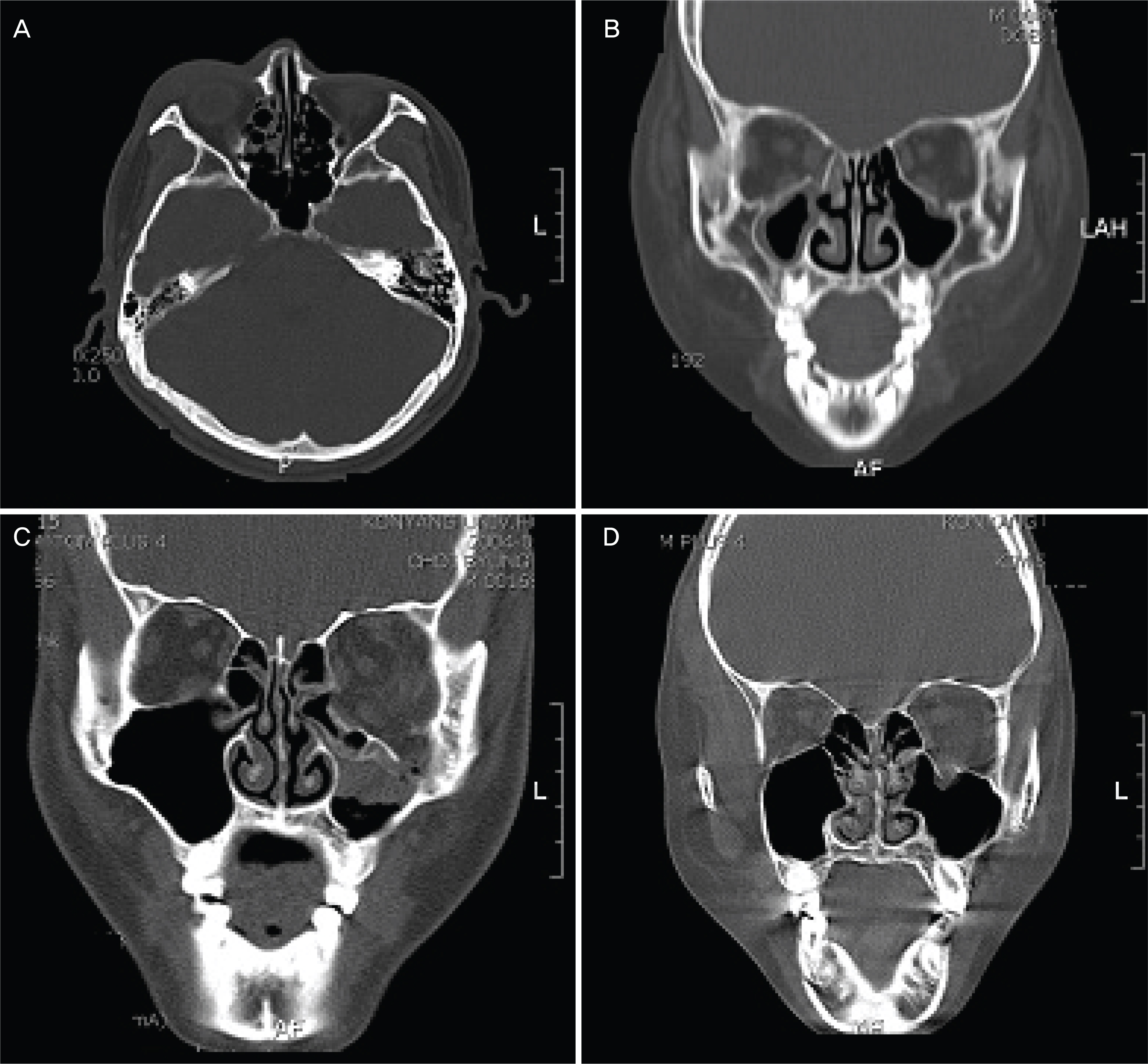
A Clinical Feature of the Patients of Orbital Wall Fracture With Diplopia
- Affiliations
-
- 1Department of Ophthalmology, Konyang University College of Medicine, Daejeon, Korea. hmseye@hanmail.net
- KMID: 2212478
- DOI: http://doi.org/10.3341/jkos.2009.50.7.969
Abstract
- PURPOSE
To analyze the clinical features of orbital wall fracture with diplopia between the surgical treatment group and the conservative treatment group. METHODS: The study comprised of 109 eyes of 109 patients with orbital wall fracture and diplopia. The patients were divided into two groups: the surgical treatment group (59 cases) and the conservative treatment group (50 cases). The groups were analyzed retrospectively according to age, gender, cause, CT, the period and severity of diplopia, and enophthalmos with time. RESULTS: In the conservative treatment group, 38 cases (64.4%) had medial wall fracture, and the average fracture size was 26alpha of the inferior wall and 33% of the medial wall. In addition, at the first visit, the patients showed diplopia within 45.5 degrees, and diplopia disappeared completely within 17 days on average (57 cases, 96.6%). In the group that underwent the reconstruction of orbital wall fracture, 27 cases (54.0%) had inferior wall fracture, and the average fracture size was 41% of the inferior wall and 35% of the medial wall. Additionally, in the first visit, the patients showed diplopia within 20.3 degrees. The muscle incarceration occurred in 12 cases (24%). In the surgical treatment group, diplopia disappeared completely within 30 days on average (45 cases, 90.0%). CONCLUSION: In the group of conservative treatment, they showed diplopia within 45.5 degrees at the first visit. Diplopia disappeared completely within 17 days on average (57 cases, 96.6%). In the group of surgical treatment, they showed diplopia within 20.3 degrees at the first visit. Diplopia disappeared completely within 30 days on average (45 cases, 90.0%).
Figure
Cited by 4 articles
-
Factors Affecting Persistent Diplopia after Surgical Repair of Isolated Inferior Orbital Wall Fracture
Joseph Kim, Sung Mo Kang
J Korean Ophthalmol Soc. 2019;60(2):181-186. doi: 10.3341/jkos.2019.60.2.181.Clinical Manifestations and Computed Tomography Findings of Trapdoor Type Medial Orbital Wall Blowout Fracture
Sung Ha Hwang, Su jin Park, Mijung Chi
J Korean Ophthalmol Soc. 2020;61(2):117-124. doi: 10.3341/jkos.2020.61.2.117.Clinical Features for Patients Presenting with Diplopia
Min Seok Kim, Jin Choi, Jung Hoon Kim, Jae Suk Kim, Joo Hwa Lee
J Korean Ophthalmol Soc. 2013;54(11):1772-1777. doi: 10.3341/jkos.2013.54.11.1772.Comparison of Diplopia and Ocular Torsion Rate in Blow-Out Fracture Patients
Kyoung Lae Kim, Sung Pyo Park, Hyoung Kyun Kim
J Korean Ophthalmol Soc. 2015;56(2):162-167. doi: 10.3341/jkos.2015.56.2.162.
Reference
-
References
1. Hosal BM, Beatty RL. Diplopia and enopthalmos after surgical repair of blowout fracture. Orbit. 2002; 21:27–33.2. Putterman AM, Stevens T, Urist MJ. Nonsurgical management of blowout fractures of the orbital floor. Am J Ophthalmol. 1974; 77:232–9.
Article3. Gilbard SM, Mafee MF, Lagouros PA, Langer BG. Orbital blowout fractures. The prognostic significance of computed tomography. Ophthalmology. 1985; 92:1523–8.
Article4. Pearl RM. Treatment of enopthalmos. Clin Plast Surg. 1992; 19:99–111.5. Iliff NT. The opthalmic implications of the correction of late enophthalmos following severe midfacial trauma. Trans Am Ophthalmol Soc. 1991; 89:477–548.6. Hwang JH, Kwag MS. Residual diplopia and enophthalmos after reconstruction of rbital wall fractures. J Korean Ophthalmol Soc. 2003; 44:1959–65.7. Kim HY, Kim YI, Won IK. Clinical analysis of blowout fracture with ocualr motion limitation: comparison of surgical and conservative treatment. J Korean Ophthalmol Soc. 1999; 40:632–8.8. Kim SK, Chang HK. The clinical study of treatment of blowout fracture. J Korean Ophthalmol Soc. 1995; 36:1629–35.9. Kroll M, Wolper J. Orbital blowout fracture. Am J Ophthalmol. 1967; 64:1169–72.10. Yang PJ, Chi NC, Choi GJ. Comparison of sugical outcome between early and delayed repair of orbital wall fracture. J Korean Ophthalmol Soc. 2004; 44:1278–84.11. Haug RH, Van Sickels JE, Jenkins WS. Demographics and treatment options for orbital roof fractures. Oral Surg Oral Med Oral Pathol Oral Radiol Endod. 2002; 93:238–46.
Article12. Smith B, Regan WF Jr. Blowout fracture of the orbit. Mechanism and correction of internal orbital fracture. Am J Ophthalmol. 1957; 44:739–9.13. Putterman AM. Management of blow out fracture of the orbital floor: The conservative approach. Surv Ophthalmol. 1991; 35:292–8.14. Millman AL, Della Rocca RC, Spector S, et al. Steroid and orbital blowout fractures, A new systematic concept in medical management and surgical decision-making. Adv Ophthalmic Plast Reconstr Surg. 1987; 6:291–300.15. Koorneef L. Current concepts on the management of orbital blowout fracture. Ann Plast Surg. 1982; 9:195–200.16. Everhard-Halm YS, Koornneef L, Zonneveld FW. Conservative therapy frequently indicated in blowout fractures of the orbit. Ned Tijdschr Geneeskd. 1991; 135:1226–8.17. Wilkins RB, Havins WE. Current treatment of blowout fractures. Ophthalmology. 1982; 89:464–6.
Article


