A Case of Cataract Operation Using Iris Retractor in Congenital Microcoria
- Affiliations
-
- 1Department of Ophthalmology, KyungHee University College of Medicine, Seoul, Korea. khjinmd@khmc.or.kr
- KMID: 2212265
- DOI: http://doi.org/10.3341/jkos.2009.50.4.618
Abstract
-
PURPOSE: To report an case regarding phacoemulsification and intraocularlens (IOL) implantation using an iris retractor in a congenital microcoria patient.
CASE SUMMARY
A 53-year-old male patient visited the office with a complaint of gradual decrease of visual acuity. The patient's best corrected visual acuity (BCVA) was 0.3, and his refractive index could not be measured, as a very small pupil was observed in both eyes. After applying tropicamide and phenylephrine four times, the refractive index of both eyes remained immeasurable. In addition, on slit-lamp biomicroscopic examination, a nucleosclerotic cataract was observed in both eyes. Any signs of increased intraocular pressure or gonioscopic findings seen in glaucomatous patients was not detected in either eyes. Using an ultrasound biomicroscope and Pentacam, the pupil diameter were 0.31 mm in both eyes, and changed to 0.92 mm in the right eye, and 1.0 mm in the left eye, after applying mydriatics. Phacoemulsification and IOL implantation were performed one month apart from each other, using an iris retractor. On the follow-up examination 2 to 3 months postoperatively, BCVA was 0.7 in the right eye, and 1.0 in the left eye. After the operation, the pupil diameter was not changed before and after mydriasis, and measured 2.53 mm in the right eye, and 2.83 mm in the left eye.
CONCLUSIONS
The authors have reported a case regarding visual acuity that improved in a congenital microcoria patient performing a phacoemulsification and IOL implantation operation using an iris retractor.
MeSH Terms
Figure
-

Figure 1. Anterior segment photograph of congenital microcoria. (A) Before topical infilatration of mydriatics (right eye). (B) Before topical infiltration of mydriatics (left eye). (C) After topical infilatration of mydriatics (right eye). (D) After topical infiltration of mydriatics (left eye).

Figure 2. Pedigree of congenital microcoria. White square=non-affected male. White circle=non-affected female. Black square=affected male. Black circle=affected female. The proband is indicated by black arrow. Question mark=un-examined person. II-1 was excluded due to iris injury and corneal neovascularization. Diagonal line=dead person.

Figure 3. A ultrasound biomicroscopy (UBM) of both the eyes shows shallow anterior chamber, convex iris, and constricted pupil. White arrows indicate pupil. (A) Physiologic state of microcoria shows severe constricted pupil (0.31 mm in diameter) in the right eye. (B) After topical mydriatic infiltration, the constricted pupil is still seen in the right eye (pupil diameter 0.92 mm) (C) Physiologic state of microcoria shows severe constricted pupil (pupil diameter 0.31 mm) in the left eye. (D) After topical mydriatic infiltration, constricted pupil is still seen in the left eye (pupil diameter 1.0 mm).
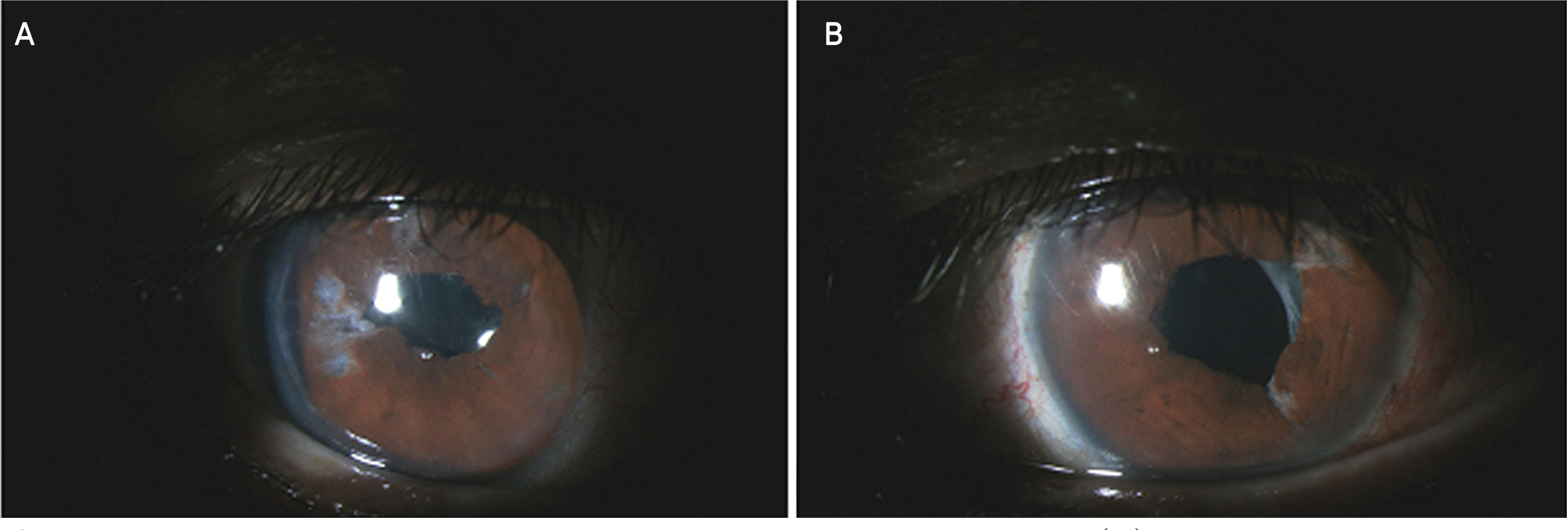
Figure 4. Postoperative anterior segment photograph of congenital microcoria. (A) Mid-dilated irregular-shaped pupil of the right eye is seen due to iris sphincter rupture and iris injury. Slightly upward displaced pupil of the right eye is seen. (B) Mid-dilated irregular-shaped pupil of the left eye is seen due to iris sphincter rupture and iris injury.
Reference
-
References
1. Pietropaolo A, Corvino C, DeBlasi A, Calabrò F. Congenital microcoria: Case report and histologic study. J Pediatr Ophthalmol Strabismus. 1998; 35:125–7.2. Toulemont PJ, Urvoy M, Coscas G, et al. Association of congenital microcoria with myopia and glaucoma. A study of 23 patients with congenital microcoria. Ophthalmology. 1995; 102:193–8.3. Rouillac C, Roche O, Marchant D, et al. Mapping of congenital microcoria locus to 13q31-q32. Am J Hum Genet. 1998; 62:1117–22.4. Zenker M, Tralau T, Lennert T, et al. Congenital nephrosis, mesangial sclerosis, and distinct eye abnormalities with microcoria: An autosomal recessive syndrome. Am J Med Genet A. 2004; 130:138–45.
Article5. Ramprasad VL, Sripriya S, Ronnie G, et al. Genetic homogeneity for inherited congenital microcoria loci in an asian indian pedigree. Mol Vis. 2005; 11:934–40.6. Kong IH. Congenital miosis: 4 cases in a family. J Korean Ophthalmol Soc. 1967; 8:31–2.7. Bremner FD, Houlden H, Smith SE. Genotypic and phenotypic heterogeneity in familial microcoria. Br J Ophthalmol. 2004; 88:469–73.
Article8. Tawara A, Itou K, Kubota T, et al. Congenital microcoria associated with lateonset developmental glaucoma. J Glaucoma. 2005; 14:409–13.
Article9. Vila-coro AA, Mazow ML, Holladay JT, et al. Congenital Idiopathic microcoria. Am J Ophthalmol. 1989; 15:439–40.
Article
