Leiomyosarcoma of the Inferior Vena Cava Mimicking Right Adrenal Tumor
- Affiliations
-
- 1Department of Surgery, Cheongju St. Mary's Hospital, Cheongju, Korea. gsurgeonl@naver.com
- 2Department of Pathology, Cheongju St. Mary's Hospital, Cheongju, Korea.
- 3Center for Health Promotion, Samsung Medical Center, Seoul, Korea.
- KMID: 2211961
- DOI: http://doi.org/10.4174/jkss.2010.78.5.330
Abstract
- The most frequent tumor arising from retroperitoneum is sarcoma. Most sarcomas of retroperitoneal origin have no symptoms and comprise 15% of all sarcomas. However, they can grow so great as to cause pain, which implies the possibility of invasion to adjacent organs. Moreover, if its location is between right adrenal gland and inferior vena cava (IVC) ambiguity of its origin can arise. Leiomyosarcoma of IVC is so rare that it can be seen to mimic right adrenal tumor. This 56-year-old female patient with back pain since approximately 4 months prior was transferred to our hospital via local clinic. We performed radical resection of tumor including segmental resection of IVC. Final diagnosis was leiomyosarcoma of IVC. We report this case with a review of literature.
Keyword
MeSH Terms
Figure
-
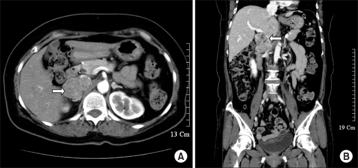
Fig. 1 Abdominal CT shows a large lobulated mass with partial invasion of inferior vena cava (IVC) and heterogeneous delayed enhancement between IVC and right adrenal gland.
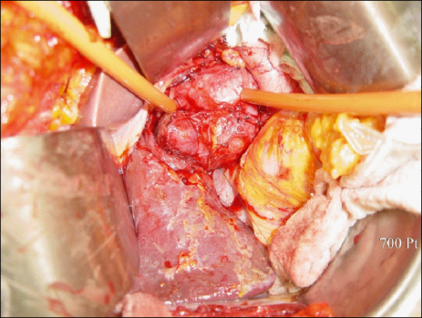
Fig. 2 Intra-operative finding shows cannulated inferior vena cava (IVC) between Infrahepatic portion and just above portion of renal vein with vascular tourniquets, which the tumor about 7 cm in diameter surrounds from the right side of IVC to adjacent to right adrenal gland.

Fig. 3 (A) Cut surface of specimen shows grayish and hard shape with mucoid change and necrosis. (B) The slide shows intravascular proliferation of atypical spindle cells (H&E stain, ×100). (C) The slide shows leiomyosarcoma showing many atypical mitoses and nuclear anaplasia (H&E stain, ×400).
Reference
-
1. Stoeckle E, Coindre JM, Bonvalot S, Kantor G, Terrier P, Bonichon F, et al. Prognostic factors in retroperitoneal sarcoma: a multivariate analysis of a series of 165 patients of the French Cancer Center Federation Sarcoma Group. Cancer. 2001. 92:359–368.2. Mendenhall WM, Zlotecki RA, Hochwald SN, Hemming AW, Grobmyer SR, Cance WG. Retroperitoneal soft tissue sarcoma. Cancer. 2005. 104:669–675.3. Lewis JJ, Leung D, Woodruff JM, Brennan MF. Retroperitoneal soft-tissue sarcoma: analysis of 500 patients treated and followed at a single institution. Ann Surg. 1998. 228:355–365.4. Dew J, Hansen K, Hammon J, McCoy T, Levine EA, Shen P. Leiomyosarcoma of the inferior vena cava: surgical management and clinical results. Am Surg. 2005. 71:497–501.5. Mingoli A, Cavallaro A, Sapienza P, Di Marzo L, Feldhaus RJ, Cavallari N. International registry of inferior vena cava leiomyosarcoma: analysis of a world series on 218 patients. Anticancer Res. 1996. 16:3201–3205.6. Burke AP, Virmani R. Sarcomas of the great vessels. A clinicopathologic study. Cancer. 1993. 71:1761–1773.7. Kieffer E, Alaoui M, Piette JC, Cacoub P, Chiche L. Leiomyosarcoma of the inferior vena cava: experience in 22 cases. Ann Surg. 2006. 244:289–295.8. Hollenbeck ST, Grobmyer SR, Kent KC, Brennan MF. Surgical treatment and outcomes of patients with primary inferior vena cava leiomyosarcoma. J Am Coll Surg. 2003. 197:575–579.9. Illuminati G, Calio FG, D'Urso A, Giacobbi D, Papaspyropoulos V, Ceccanei G. Prosthetic replacement of the infrahepatic inferior vena cava for leiomyosarcoma. Arch Surg. 2006. 141:919–924.
