Two Cases of Occult Macular Dystrophy in a Family
- Affiliations
-
- 1Department of Ophthalmology, Seoul National University College of Medicine Seoul Artificial Eye Center, Seoul National University Hospital Clinical Research Institute, Seoul, Korea. jiani4@snu.ac.kr
- 2Department of Ophthalmology, Seoul National University Bundang Hospital, Gyeonggi, Korea.
- KMID: 2211720
- DOI: http://doi.org/10.3341/jkos.2008.49.5.858
Abstract
-
PURPOSE: We report two familial cases of occult macular dystrophy (OMD) presenting with a progressive decrease in central vision.
CASE SUMMARY
Both patients exhibited a normal ophthalmologic examination including slit lamp biomicroscope, fundus examination, fluorescein angiography, and full-field electroretinogram. However, there were central visual field defects on a Humphrey static visual field test (C 24-2) and abnormal multifocal electroretinogram (mfERG) findings affecting the central portion of the test field. Foveal thinning was also observed by optical coherence tomography images in 1 case. These findings are consistent with the clinical characteristics of occult macular dystrophy, and close observation was recommended.
CONCLUSIONS
OMD is a disease characterized by a reduction in central visual acuity without visible fundus abnormalities and full field ERG. It may be misdiagnosed as optic nerve disease, a central nervous system problem, non-organic visual disorder or malingering; therefore, mfERG is essential for the diagnosis of this rare type of macular dystrophy.
MeSH Terms
Figure
-

Figure 1. Ophthalmoscopic (top) and fluorescein angiographic (bottom) findings in the right (shown left) and left (shown right) eyes of patient 1. There is no detectable abnormality at the disc and around the macular regions of either eye.
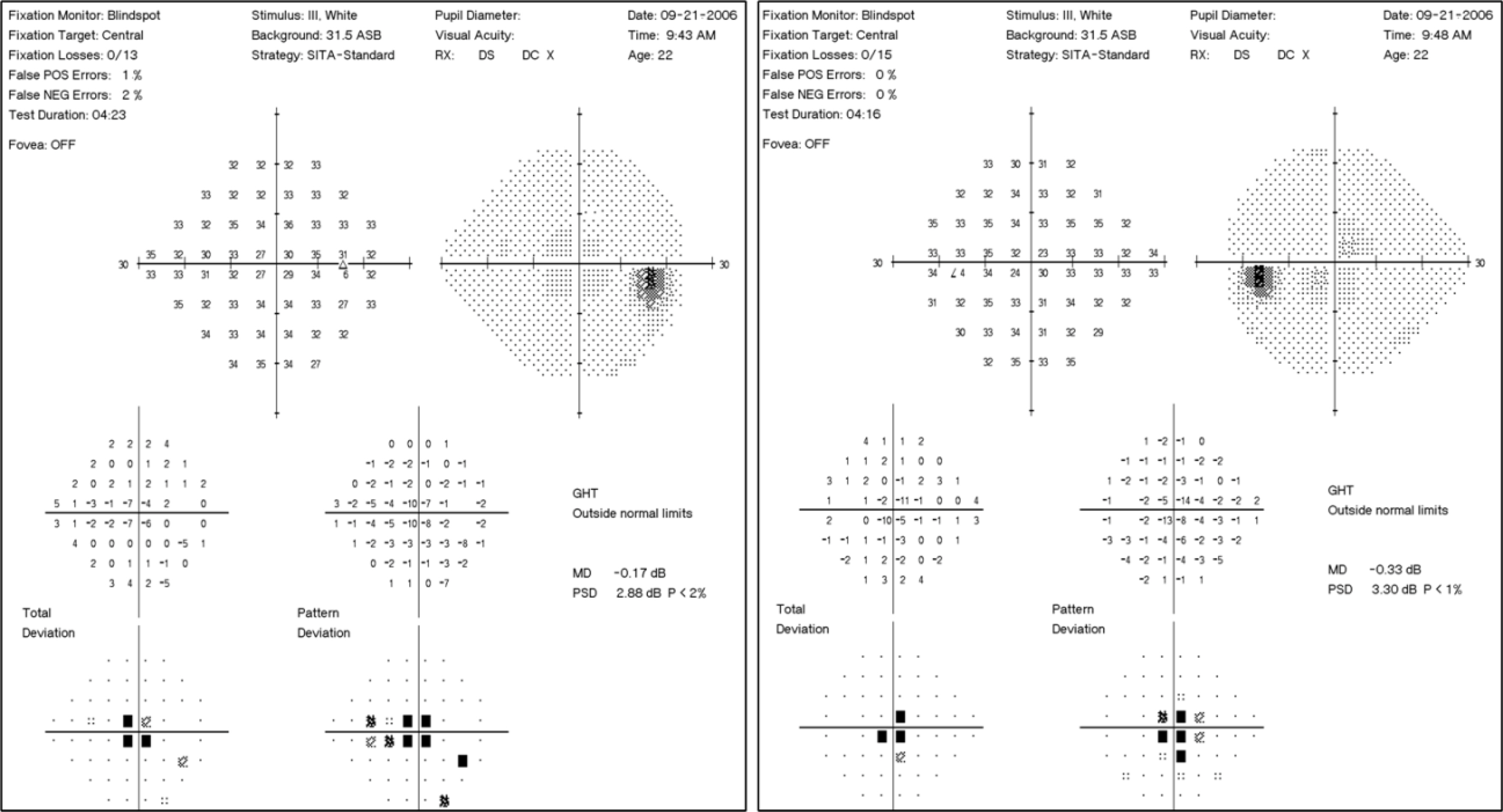
Figure 2. Humphrey C24-2 perimetry of the right (shown left) and left (shown right) eyes of patient 1. There is a central scotoma in both eyes.
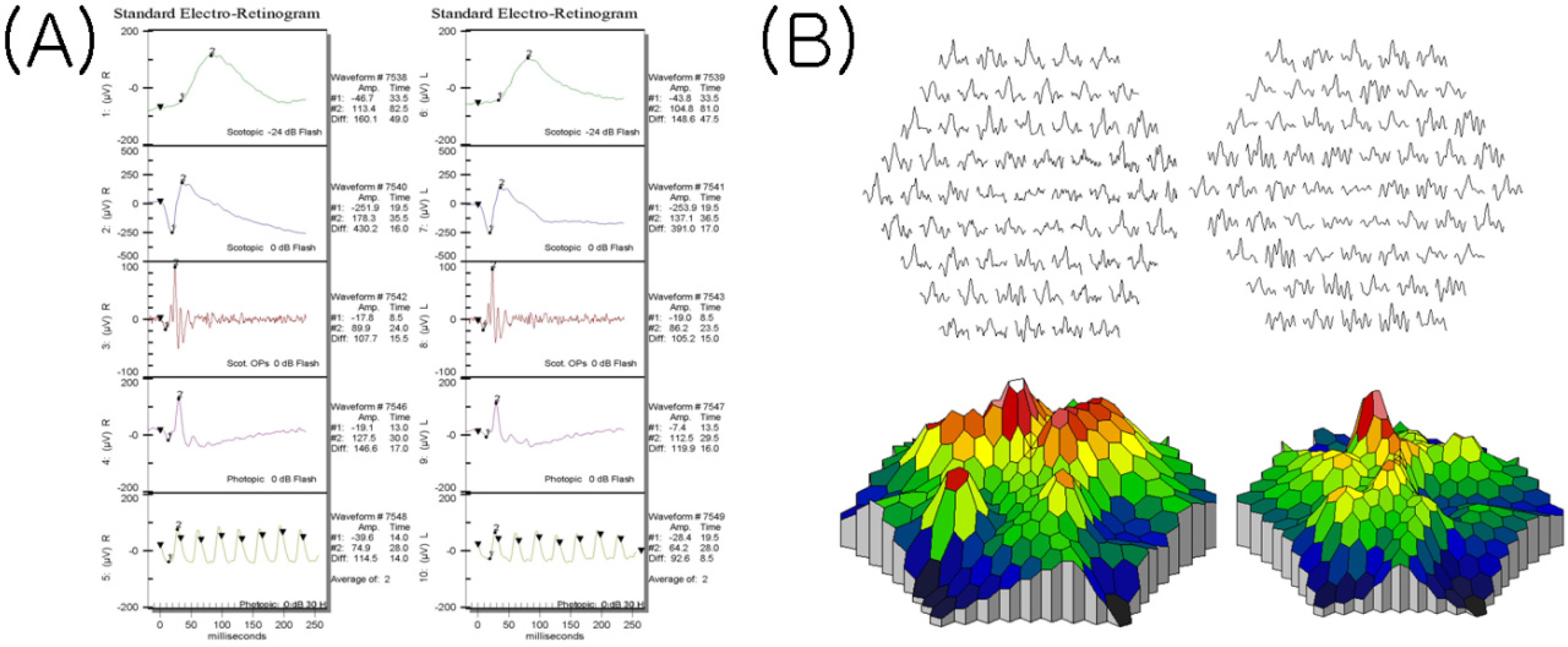
Figure 3. (A) The photopic and scotopic full field ERG was normal in both eyes of patient 1. (B) The multifocal electroretinographic responses from the central areas are attenuated in the right (shown left) and left (shown right) eyes.

Figure 4. The 3 mm horizontal scans of the optical coherence tomography images obtained from patient 1 demonstrating foveal thickness of 114 µm in the right eye (left), 108 µm in the left eye (right).
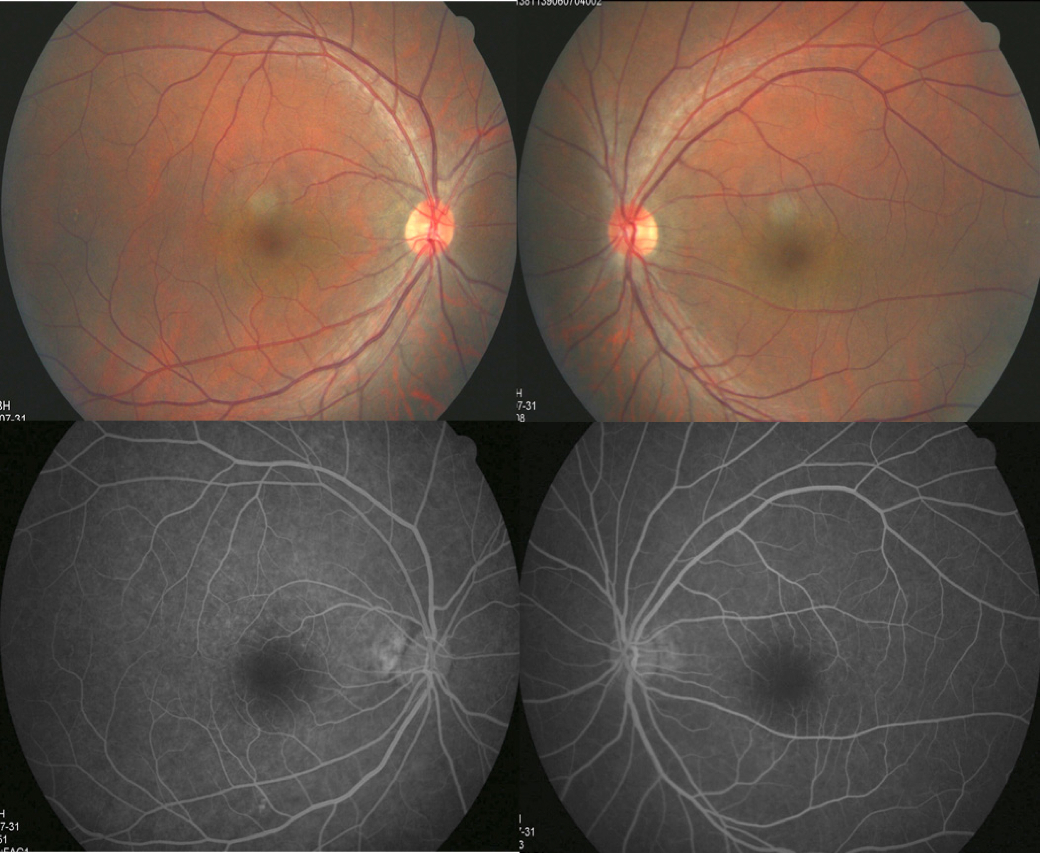
Figure 5. Ophthalmoscopic (top) and fluorescein angiographic (bottom) findings in the right (shown left) and left (shown right) eyes of patient 2. There is no detectable abnormality at the disc and around the macular regions of either eye.
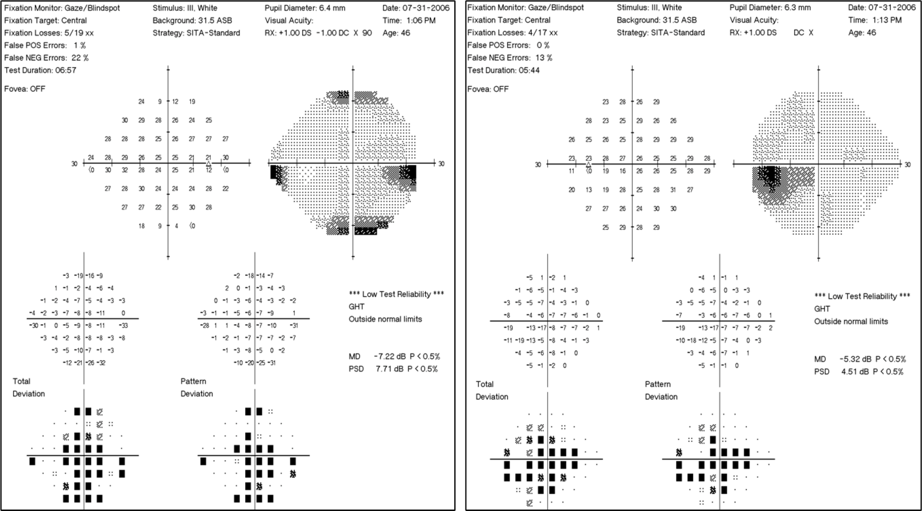
Figure 6. Humphrey C24-2 perimetry of right (shown left) and left (shown right) eyes of patient 2. There is a large central field defect in both eyes.
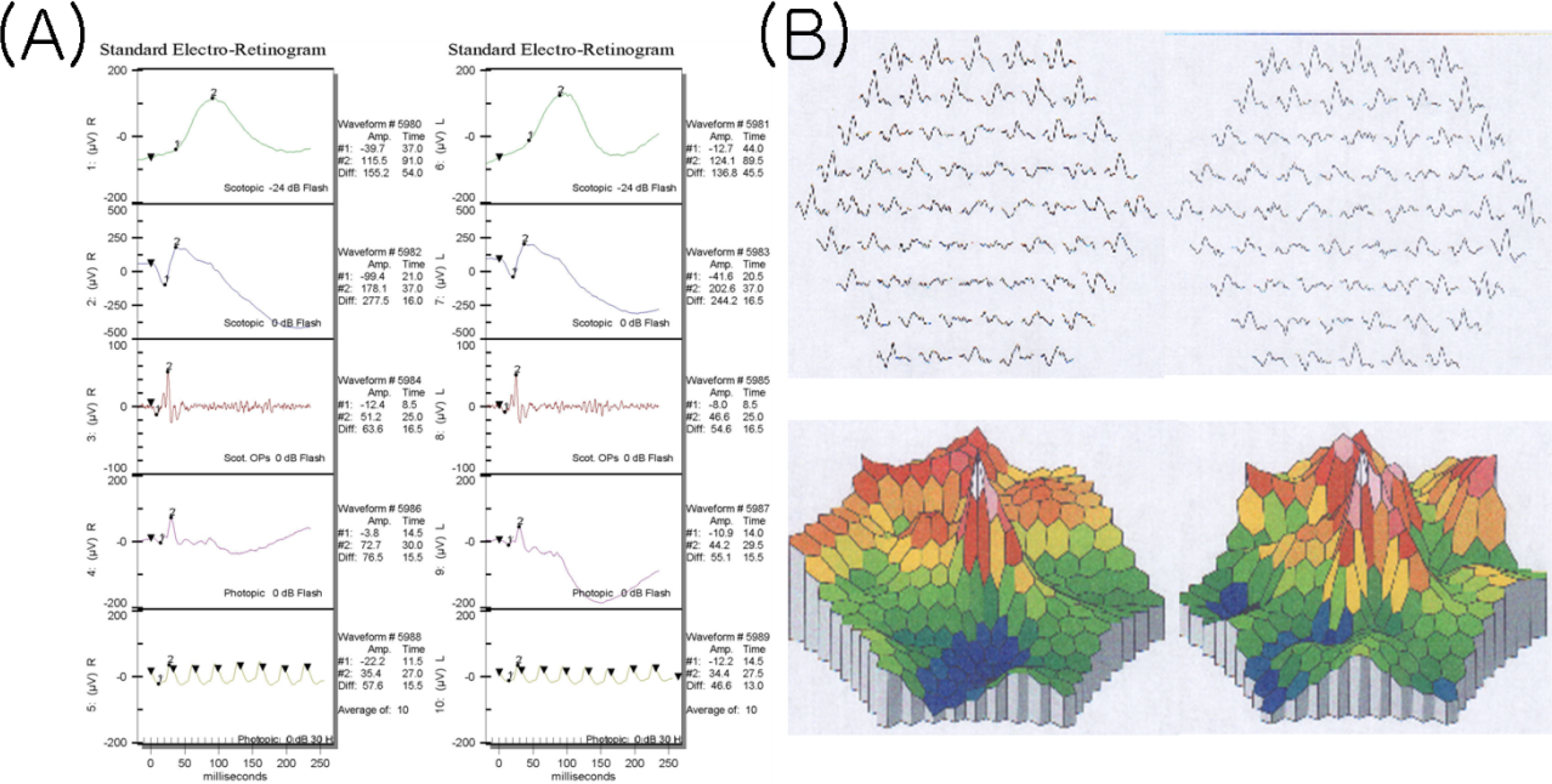
Figure 7. (A) The photopic and scotopic full field ERG showed mildly decreased amplitude and normal implicit time in both eyes of patient 2. (B) The multifocal electroretinographic responses from the central areas are attenuated in the right (shown left) and left (shown right) eyes.
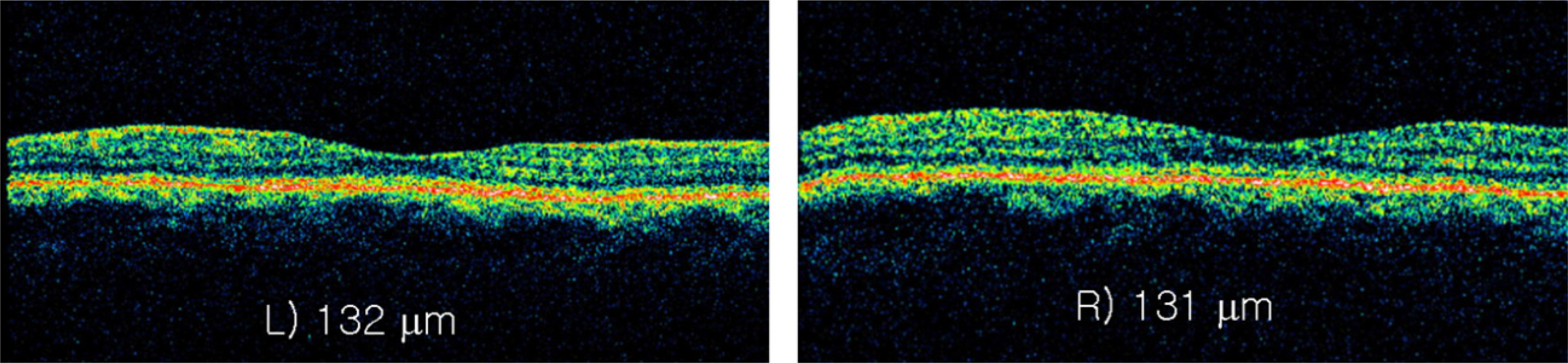
Figure 8. The 3 mm horizontal scans of the optical coherence tomography images obtained from patient 2 demonstrating foveal thickness of 131 µm in the right eye (shown left), 132 µm in the left eye (shown right).
Reference
-
References
1. Miyake Y, Ichikawa K, Shiose Y, Kawase Y. Hereditary macular dystrophy without visible fundus abnormality. Am J Ophthalmol. 1989; 108:292–9.
Article2. Wildberger H, Niemeyer G, Junghardt A. Multifocal electroretinogram (mfERG) in a family with occult macular dystrophy. Klin Monatsbl Augenheilkd. 2003; 220:111–5.3. Lyons JS. Non-familial occult macular dystrophy. Doc Ophthalmol. 2005; 111:49–56.
Article4. Miyake Y, Horiguchi M, Tomita N, et al. Occult macular dystrophy. Am J Ophthalmol. 1996; 122:644–53.
Article5. Sandberg MA, Brockhurst RJ, Gaudio AR, Berson EL. The association between visual acuity and central retinal thickness in retinitis pigmentosa. Invest Ophthalmol Vis Sci. 2005; 46:3349–54.
Article6. Kondo M, Ueno S, Piao CH, et al. Occult macular dystrophy in an 11 year old boy. Br J Ophthalmol. 2004; 88:1602–3.
Article7. Piao CH, Kondo M, Tanikawa A, et al. Multifocal electroretinogram in occult macular dystrophy. Invest Ophthalmol Vis Sci. 2000; 41:513–7.8. Kondo M, Ito Y, Ueno S, et al. Foveal thickness in occult macular dystrophy. Am J Ophthalmol. 2003; 135:725–8.
Article9. Brockhurst RJ, Sandberg MA. Optical coherence tomography findings in occult macular dystrophy. Am J Ophthalmol. 2007; 143:516–8.
Article10. Ergun E, Hermann B, Wirtitsch M, et al. Assessment of central visual function in Stargardt disease/fundus flavimaculatus with ultrahigh-resolution optical coherence tomography. Invest Ophthalmol Vis Sci. 2005; 46:310–6.
- Full Text Links
-
- Actions
-
Cited
- CITED
-
- Close
- Share
-
- Similar articles
-
- Cases of Macular Corneal Dystrophy with a Family History
- Granular Corneal Dystrophy
- A Korean Family with an Early-Onset Autosomal Dominant Macular Dystrophy Resembling North Carolina Macular Dystrophy
- A Case of Macular Dystrophy of the Cornea
- Two Cases of Oculopharyngeal Muscular Dystrophy in One Family
