J Korean Ophthalmol Soc.
2008 Apr;49(4):562-569. 10.3341/jkos.2008.49.4.562.
Clinical Characteristics of Paranasal Sinus Mucoceles Which Invade the Orbit
- Affiliations
-
- 1Department of Ophthalmology, College of Medicine, Inje University, Pusan, Korea. eyeyang@inje.ac.kr
- 2Ophthalmology Research Foundation, Inje University, Pusan, Korea.
- KMID: 2211485
- DOI: http://doi.org/10.3341/jkos.2008.49.4.562
Abstract
-
PURPOSE: We report the clinical features of paranasal sinus mucoceles with orbital extension and compare the results of external and transnasal approaches based on the rates of complications and recurrence.
METHODS
Thirty-three cases of paranasal sinus mucoceles with orbital extension diagnosed at our hospital from 2003 to 2007 were retrospectively reviewed.
RESULTS
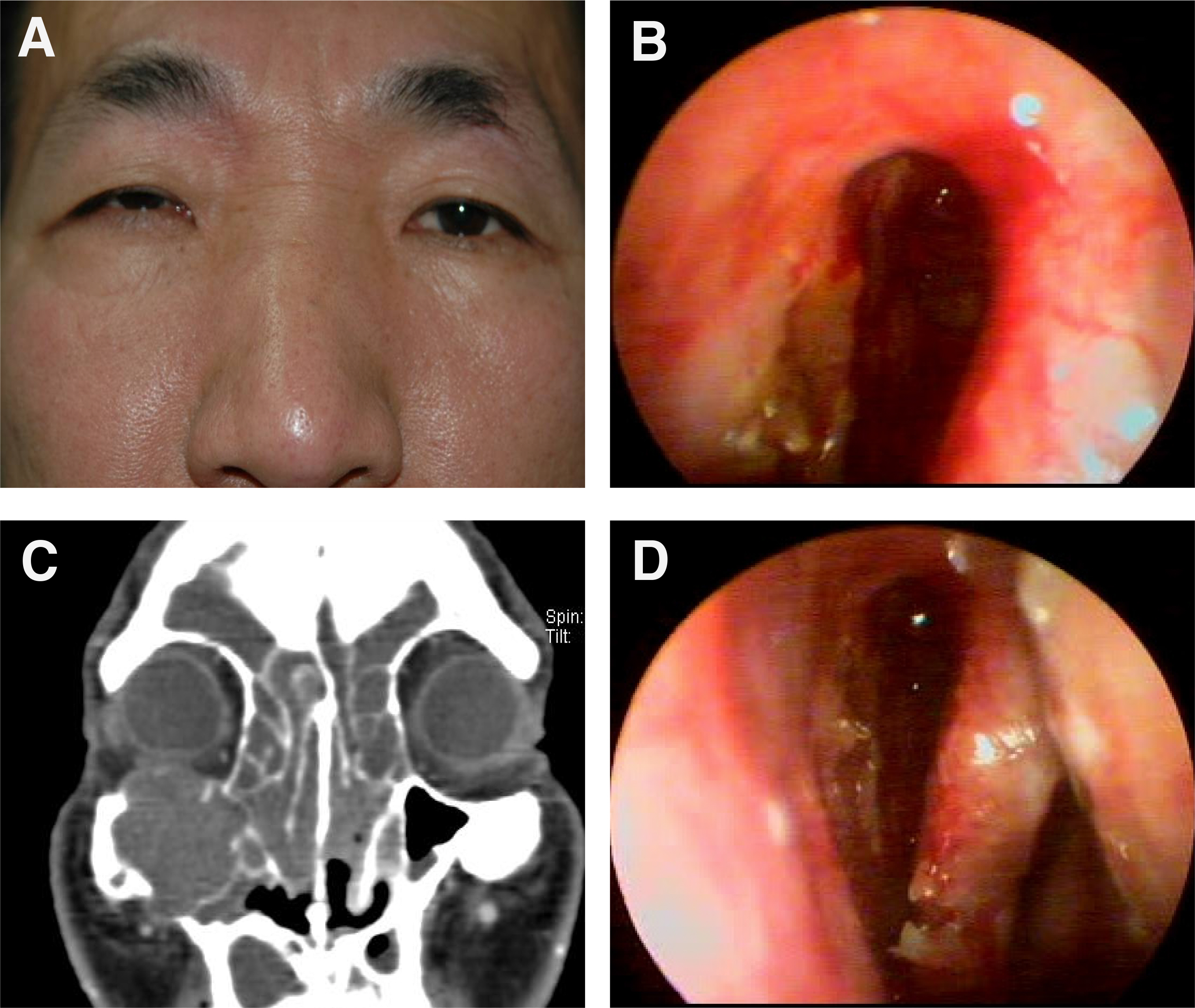
The mean age of patients was 48.6 years. The common sites of origin were the frontal, ethmoidal, frontoethmoidal sinuses, and proptosis was the most common presenting feature. Among the mucoceles of frontal and frontoethmoid sinuses, there was no difference in the rates of recurrence or complications between the two different methods.
CONCLUSIONS
Mucoceles with orbital involvement generally present with a noninfiltrating mass resulting in many ophthalmic signs and symptoms. Obliteration of the involved sinus is not recommended if there is erosion of the sinus bony wall with extension of the mucocele into the orbit. The mucosa lining the mucocele become adhered to the orbital periosteum and cannot be removed during surgery without significant risk of injury to the adjacent structures. Endoscopic sinus surgery is considered effective for paranasal sinus mucoceles with orbital involvement.
Keyword
Figure
Cited by 1 articles
-
The Effect of Endonasal Dacryocystorhinostomy in the Paranasal Mucocele Invading Nasolacrimal Duct
Gyu Chul Chung, Seung Hwan Jo, Jae Wook Yang
J Korean Ophthalmol Soc. 2018;59(3):203-208. doi: 10.3341/jkos.2018.59.3.203.
Reference
-
References
1. Beasley NJ, Jones NS. Paranasal sinus mucoceles: modern management. Am J Rhinol. 1995; 9:251–6.
Article2. Kennedy DW, Josephson JS, Zinreich SJ, et al. Endoscopic sinus surgery for mucocele: a variable alternative. Laryngoscope. 1989; 99:885–95.3. Haik BG, Amedee RG. Principles and practice of ophthalmic plastic and reconstructive surgery. 2. Philadelphia: WB Saunders;1996. p. 1014–22.4. Evans C. Aetiology and treatment of frontoethmoidal mucocele. J Laryngol Otol. 1981; 95:361–75.
Article5. Moriyama H, Nakajima T, Honda Y. Studies on mucoceles of the ethmoid and sphenoid sinuses: Analysis of 47 cases. J Laryngol Otol. 1992; 106:23–7.6. Bordley JE, Bosley WR. Mucoceles of the frontal sinus: Causes and treatment. Ann Otol Rhinol Laryngol. 1973; 82:696–702.
Article7. Van Tassel P, Lee YY, Jing BS, De Pena CA. Mucoceles of paranasal sinuses: MR imaging with CT correction. Am J Radiol. 1989; 153:407–12.8. Friedman WH. Surgery of the paranasal sinuses. 2nd ed.Philadelphia: Saunders;1991. p. 219–28.9. Toriumi DM, Berktold RE. Multiple frontoethmoid mucoceles. Ann Otol Rhinol Laryngol. 1989; 98:831–3.
Article10. Shikowitz MJ, Goldenstein MN, Stegnjajic A. Sphenoid sinus mucocele masquerading as a skull base malignancy. Laryngoscope. 1986; 96:1405–10.
Article11. Chui MC, Briant TD, Gray T, et al. Computed tomography of sphenoid sinus mucocele. J Otolaryngol. 1983; 12:263–9.12. Maisel RH, Deeb ME, Bore RC, et al. Sphenoid sinus mucoceles. Laryngoscope. 1973; 27:930–9.
Article13. Wang TJ, Liao SL, Jou JR. Clinical manifestations and management of orbital mucocele:the role of ophthalmologists. Jpn J Ophthalmol. 2005; 49:239–45.14. Moriyama H, Nakajima T, Honda Y. Studies on mucoceles of the ethmoid and sphenoid sinuses: analysis of 47 cases. J Laryngol Otol. 1992; 106:23–7.15. Sharma GD, Doeshud CF, Stern RC. Erosion of the wall of the frontal sinus caused by mucopyocele in cystic fibrosis. J Pediatr. 1994; 124:745–7.
Article16. Kim HI, Moon JI, Chung SK. A case of ethmoid sinus mucocele with transient intraocular pressure elevation. J Korean Ophthalmol Soc. 1991; 32:1015–9.17. Ormerod LD, Weber AL, Rauch SD. Ocular manifestations of maxillary sinus mucoceles. Ophthalmology. 1987; 94:1013–9.18. Stankiewiez JA. Sphenoid sinus mucocele. Arch Otolaryngol. 1989; 115:735–41.19. Lee JJ, Ha HB, Na IG, et al. A case of mucocele of the sphenoid sinus causing sudden onset of blindness and hyperprolactinemia. Korean J Otolaryngol. 1989; 39:730–3.20. Cho SH, Cho KK, Lee KS, et al. A case of sphenoid sinus mucocele with visual field defect. Korean J Neurosurg. 1992; 21:1338–42.21. Palmer BW. Unilateral exophthalmos. Arch Otolaryngol. 1965; 82:415–24.
Article22. Simon M, Tingwald F. Syndrome associated with mucocele of sphenoid sinus. Radiology. 1955; 64:538–45.23. Toriumi DM, Sykes JM, Russel EJ, et al. Sphenoethmoid mucocele with intracranial extension. Otolaryngol Head Neck Surg. 1988; 98:254–8.24. Stankiewicz MJ, Goldstein MN. Sphenoid sinus mucocele masquerading as a skull base malignancy. Laryngoscope. 1986; 96:1405–10.25. Cho EY, Choi YK, Choi WC. A case of nasal endoscopic treatment for paranasal mucocele. J Korean Ophthalmol Soc. 2004; 45:1386–91.26. East D. Mucoceles of the maxillary antrum:description, case reports and review of the literature. J Laryngol Otol. 1985; 99:49–56.27. Hejazi N, Witzmann A, Hassler W. Ocular manifestations of sphenoid mucoceles. Surg Neurol. 2001; 56:338–43.
Article28. Tunkel DE, Naclerio RM. Bilateral maxillary sinus mucoceles in an infant with cystic fibrosis. Otolaryngol Head Neck Surg. 1994; 111:116–20.
Article29. Har-EL G. Transnasal endoscopic management of fontal mucoceles. Otolaryngol Clin North Am. 2001; 34:243–51.30. Kim YM, Park YM, Koh YC. A case of mucocele of the sphenoid sinus causing complete visual loss. Korean J Otolaryngol. 1992; 35:590–5.31. Kim SS, Kang SS, Kim KS, et al. Clinical characteristics of primary paranasal sinus mucoceles and their surgical treatment outsome. Korean J Otolaryngol. 1998; 41:1436–9.32. Shin HS, Lee BJ, Kim JH, et al. Frontal sinus mucoceles:A comparison of extranasal operation and intranasal endoscopic marsupialization. Korea J Otolaryngol. 1996; 39:463–71.33. Kwon SH, Jeong WC. Endoscopic surgery for paranasal sinus mucocele. Korea J Otolaryngol. 1997; 40:1431–6.34. Wang TJ, Liao SL, Jou JR, et al. Clinical manifestations and management of orbital mucoceles: the role of ophthalmologists. Jpn J Ophthalmol. 2005; 49:239–45.
Article
- Full Text Links
- Actions
-
Cited
- CITED
-
- Close
- Share
- Similar articles
-
- Comparison of Clinical Characteristics between Primary and Secondary Paranasal Mucoceles
- A Case of Primary Septated Mucocele of Maxillary Sinus
- Mucocele in the maxillary sinus involving the orbit: A report of 2 cases
- Endoscopic Surgery for Paranasal Sinus Mucocele
- Clinical Characteristics of Primary Paranasal Sinus Mucoceles and Their Surgical Treatment Outcome


