A Rare Case of Thymic Gangliocytic Paraganglioma
- Affiliations
-
- 1Department of Pathology, Samsung Medical Center, Sungkyunkwan University School of Medicine, Seoul, Korea. hanjho@skku.edu
- 2Department of Thoracic Surgery, Samsung Medical Center, Sungkyunkwan University School of Medicine, Seoul, Korea.
- KMID: 2211385
- DOI: http://doi.org/10.4132/jptm.2015.07.15
Abstract
- No abstract available.
MeSH Terms
Figure
-
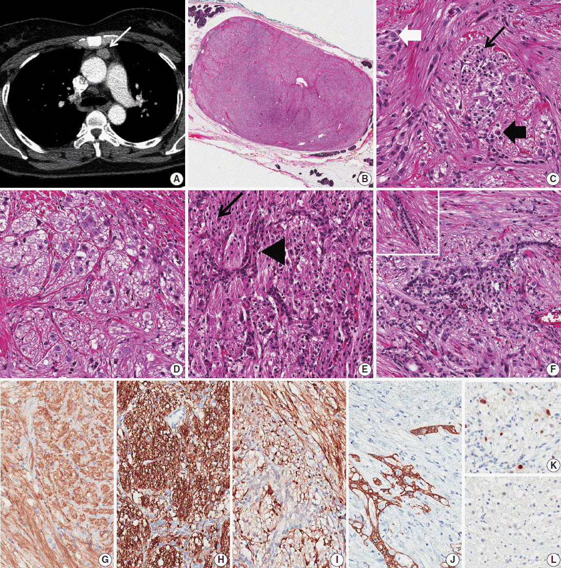
Fig. 1. Thymic gangliocytic paraganglioma. (A) Chest computed tomography reveals a well-circumscribed ovoid mass (white arrow) in the anterior mediastinum. (B) On low-power view, the mass shows an expanding border surrounded by normal thymic tissue. (C) Spindle cells show an intersecting fascicular pattern, and fibrillary cells show a vague or confluent nesting pattern (black arrow). Scattered ganglion-like cells (white thick arrow) and epithelioid cells (black thick arrow) are also noted. (D) Epithelioid cells form a vague nesting pattern. (E) Epithelial cells show a branching pattern (arrowhead) between the nests of fibrillary cells (black arrow). (F) In focal areas, the epithelial cells form compressed cords and ducts (left upper corner). (G, H) Staining with CD56 (G) and synaptophysin (H) is positive for spindle cells, ganglion-like cells, epithelioid cells, and fibrillary cells. (I) Only spindle cells are positive for S-100. Spindle cells are located adjacent to and within the nests of fibrillary cells. (J) On staining for AE1/AE3, the epithelial cells show a reticular or race-like cytoplasmic pattern. (K, L) Some of the fibrillary cells and epithelioid cells are positive (K) for progesterone receptor, but most of them are negative (L).
Reference
-
1. DeLellis RA, Lloyd RV, Heitz PU, Eng C. World Health Organization classification of tumors of pathology and genetics of tumors of endocrine organs. Lyon: IARC Press;2004. p. 162–3.2. Okubo Y, Wakayama M, Nemoto T, et al. Literature survey on epidemiology and pathology of gangliocytic paraganglioma. BMC Cancer. 2011; 11:187.
Article3. Weinrach DM, Wang KL, Blum MG, Yeldandi AV, Laskin WB. Multifocal presentation of gangliocytic paraganglioma in the mediastinum and esophagus. Hum Pathol. 2004; 35:1288–91.
Article4. de Montpréville VT, Mussot S, Gharbi N, Dartevelle P, Dulmet E. Paraganglioma with ganglioneuromatous component located in the posterior mediastinum. Ann Diagn Pathol. 2005; 9:110–4.
Article5. Weissferdt A, Kalhor N, Liu H, et al. Thymic neuroendocrine tumors (paraganglioma and carcinoid tumors): a comparative immunohistochemical study of 46 cases. Hum Pathol. 2014; 45:2463–70.
Article6. Okubo Y, Nemoto T, Wakayama M, et al. Gangliocytic paraganglioma: a multi-institutional retrospective study in Japan. BMC Cancer. 2015; 15:269.
Article7. Ogata S, Horio T, Sugiura Y, Aiko S, Aida S. Duodenal gangliocytic paraganglioma with regional lymph node metastasis and a glandular component. Pathol Int. 2011; 61:104–7.
Article
- Full Text Links
-
- Actions
-
Cited
- CITED
-
- Close
- Share
-
- Similar articles
-
- Periampullary Gangliocytic Paraganglioma Successfully Treated by Endoscopic Mucosal Resection
- First Report of a Gangliocytic Paraganglioma Arising in a Tailgut Cyst
- Gangliocytic Paraganglioma of the Duodenum
- A Case of Gangliocytic Paraganglioma of the Ampulla of Vater Presenting as Jaundice
- A Incidentally Diagnosed Duodenal Subepithelial Mass: Gangliocytic Paraganglioma Treated by Endoscopic Mucosal Resection
