J Korean Ophthalmol Soc.
2008 Mar;49(3):442-449. 10.3341/jkos.2008.49.3.442.
Treatments of Stage 1 Retinal Angiomatous Proliferation
- Affiliations
-
- 1Department of Ophthalmology, Seoul Veterans Hospital, Seoul, Korea.
- 2Department of Ophthalmology, College of Medicine, The Catholic University of Korea, Seoul, Korea. wklee@catholic.ac.kr
- KMID: 2211317
- DOI: http://doi.org/10.3341/jkos.2008.49.3.442
Abstract
-
PURPOSE: To evaluate the results of single or combined treatment methods administered to patients with stage 1 retinal angiomatous proliferation (RAP).
METHODS
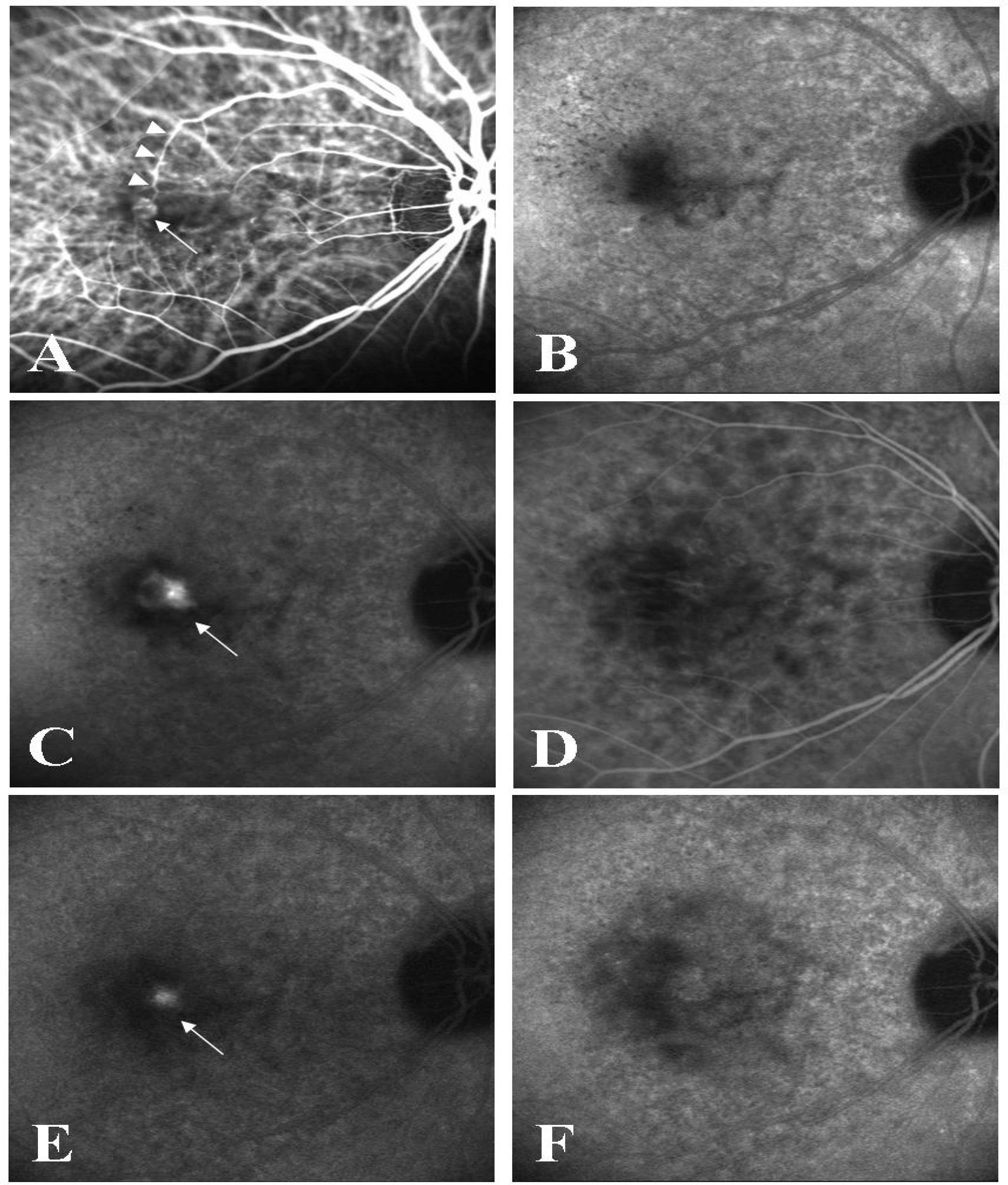
A retrospective clinical study was performed in 9 eyes of 8 patients with stage I RAP. According to fundus angiography findings taken regularly after treatment, the results were categorized as closure of stage 1, recurrence of stage 1, or progress to stage 2.
RESULTS
The mean follow-up period was 25 (14~40) months. In two eyes treated with laser photocoagulation only, the lesions progressed to stage 2. In two eyes treated with laser photocoagulation and intravitreal triamcinolone injection, the lesions also progressed to stage 2. In four eyes treated with photodynamic therapy and intravitreal triamcinolone injection, stage 1 lesions recurred in 3 eyes, and the lesion progressed to stage 2 in one eye. In one eye treated with photodynamic therapy, intravitreal triamcinolone injection, and laser photocoagulation, the stage 1 lesion recurred. Final visual acuity was stable in the case of stage 1 recurrence but was lower than the pretreatment level when the lesion progressed to stage 2.
CONCLUSIONS
With either single or combined therapy, temporary closure of the stage 1 RAP lesion was possible, but complete closure could not be achieved for a long period because the lesion either recurred or progressed.
Keyword
MeSH Terms
Figure
Cited by 2 articles
-
Twelve-Month Outcomes of Intravitreal Anti-Vascular Endothelial Growth Factor Therapy for Retinal Angiomatous Proliferation
Deok Bae Kim, Jae Hui Kim, Seong Hun Jeong, Tae Gon Lee, Jong Woo Kim, Chul Gu Kim, Sung Won Cho, Dong Won Lee, Jung I1 Han
J Korean Ophthalmol Soc. 2013;54(11):1700-1707. doi: 10.3341/jkos.2013.54.11.1700.Comparison of Optical Coherence Tomography Characteristics among Three Subtypes of Exudative Age-related Macular Degeneration
Soh-Eun Ahn, Dong Seob Ahn, Heon Yang, Hee Seong Yoon
J Korean Ophthalmol Soc. 2016;57(7):1093-1101. doi: 10.3341/jkos.2016.57.7.1093.
Reference
-
References
1. Hartnett ME, Weiter JJ, Staurenghi G, Elsner AE. Deep retinal vascular anomalous complexes in advanced age-related macular degeneration. Ophthalmology. 1996; 103:2042–53.
Article2. Kuhn D, Meunier I, Soubrane G, Coscas G. Imaging of choroidal anastomoses in vascularized retinal pigment epithelium detachments. Arch Ophthalmol. 1995; 113:1392–8.3. Yannuzzi LA, Negrao S, Ilda T, et al. Retinal angiomatous proliferation in age-related macular degeneration. Retina. 2001; 21:416–34.
Article4. Gass JD, Agarwal A, Lavina AM, et al. Focal inner retinal hemorrhages in patients with drusen: an early sign of occult choroidal neovascularization and chorioretinal anastomosis. Retina. 2003; 23:741–51.5. Axer-Siegel R, Bourla D, Priel E, et al. Angiographic and flow patterns of retinal choroidal anastomoses in age-related macular degeneration with occult choroidal neovascularization. Ophthalmology. 2002; 109:1726–36.
Article6. Lee PY, Lee WK. Progression of retinal angiomatous proliferation after surgical ablation. J Korean Ophthalmol Soc. 2006; 47:1523–32.7. Byeon SH, Hong JP, Lee H, et al. Photodynamic therapy results of retinal angiomatous proliferation with pigment epithelial detachment in age-related macular degeneration. J Korean Ophthalmol Soc. 2006; 47:1410–6.8. Bottoni F, Massacesi A, Cigada M, et al. Treatment of retinal angiomatous proliferation in age-related macular degeneration: a series of 104 cases of retinal angiomatous proliferation. Arch Ophthalmol. 2005; 123:1644–50.9. Boscia F, Furino C, Sborgia L, et al. Photodynamic therapy for retinal angiomatous proliferation and pigment epithelium detachment. Am J Ophthalmol. 2004; 138:1077–9.10. Nakata M, Yuzawa M, kawamura A, Shimada H. Combining surgical ablation of retinal inflow and outflow vessels with photodynamic therapy for retinal angiomatous proliferation. Am J Ophthalmol. 2006; 141:968–70.
Article11. Bottoni F, Romano M, Massacesi A, Bergamini F. Remodeling of the vascular channels in retinal angiomatous proliferations treated with intravitreal triamcinolone acetonide and photodynamic therapy. Graefes Arch Clin Exp Ophthalmol. 2006; 244:1528–33.
Article12. Krieglstein TR, Kampik A, Ulbig M. Intravitreal triamcinolone and laser photocoagulation for retinal angiomatous proliferation. Br J Ophthalmol. 2006; 90:1357–60.
Article13. Conti SM, Kertes PJ. The use of intravitreal corticosteroids, evidence-based and otherwise. Curr Opin Ophthalmol. 2006; 17:235–44.
Article14. Meyerle CB, Freund KB, Iturralde D, et al. Intravitreal bevacizumab (Avastin) for retinal angiomatous proliferation. Retina. 2007; 27:451–7.
Article
- Full Text Links
- Actions
-
Cited
- CITED
-
- Close
- Share
- Similar articles
-
- Progression of Retinal Angiomatous Proliferation after Surgical Ablation
- Retinal Angiomatous Proliferation
- Photodynamic Therapy Results of Retinal Angiomatous Proliferation with Pigmented Epithelial Detachment in Age-related Macular Degeneration
- Characteristic Findings of Optical Coherence Tomography in Retinal Angiomatous Proliferation
- Retinal Angiomatous Proliferation and Intravitreal Bevacizumab Injection


