J Korean Surg Soc.
2009 Aug;77(2):149-152. 10.4174/jkss.2009.77.2.149.
Recurrence after Repair of Primary Acquired Grynfeltt Hernia
- Affiliations
-
- 1Department of Surgery, Chonnam National University Medical School, Gwangju, Korea. wkafyddl@hanmail.net
- KMID: 2211296
- DOI: http://doi.org/10.4174/jkss.2009.77.2.149
Abstract
- Lumbar hernias are rare posterolateral abdominal wall defects. There are two types of lumbar hernia. One is a superior lumbar hernia through the deep superior orifice (Grynfeltt triangle), and the other is a lower lumbar hernia through the superficial lower orifice (Petit triangle). A lumbar hernia is often misdiagnosed as a lipoma, so a cautious clinical examination is very important. Reports of recurrent lumbar hernia are extremely rare in the literature. We experienced a case of recurrence in an acquired primary lumbar hernia in a 71-year-old male who had undergone mesh-plug herniorrhaphy. The hernia orifice was 1 cm in diameter and exhibited a fibrous smooth margin. Hernia repair using 3-D mesh was performed. The patient had uncomplicated postoperative course and was discharged one day after the operation.
Figure
-
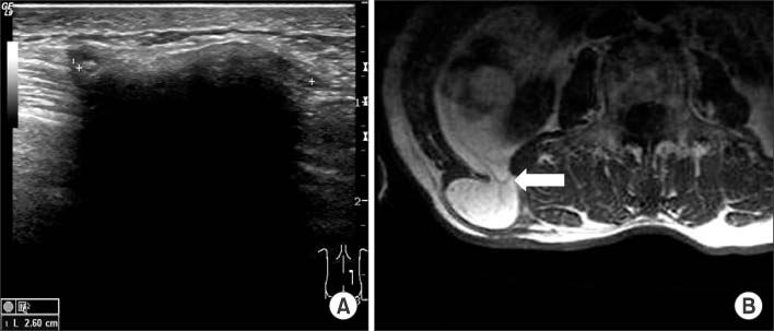
Fig. 1 Radiologic findings. (A) Ultrasonogram shows a 2.6 cm sized movable mass with marked posterior acoustic shadow. (B) Abdominal MRI reveals a 3.7×2.6×6.3 cm sized herniated retroperitoneal fat through the defect of transversalis fascia (arrow) at the level of L2~L3.

Fig. 2 Operative findings. (A) After skin incision, a displaced mesh plug is exposed. (B) The size of facial defect is about 1 cm of diameter. (C) Onlay of Prolene* 3D patch® (Ethicon, Somerville, NJ, USA) covered more than 3 cm from the margin of facial defect in all directions.
Reference
-
1. Skrekas G, Stafyla VK, Papalois VE. A Grynfeltt hernia: report of a case. Hernia. 2005. 9:188–191.2. Heniford BT, Iannitti DA, Gagner M. Laparoscopic inferior and superior lumbar hernia repair. Arch Surg. 1997. 132:1141–1144.3. Le Neel JC, Sartre JY, Borde L, Guiberteau B, Bourseau JC. Lumbar hernias in adults. Apropos of 4 cases and review of the literature. J Chir (Paris). 1993. 130:397–402.4. Armstrong O, Hamel A, Grignon B, NDoye JM, Hamel O, Robert R, et al. Lumbar hernia: anatomical basis and clinical aspects. Surg Radiol Anat. 2008. 30:533–537. discussion 609-10.5. Loukas M, El-Zammar D, Shoja MM, Tubbs RS, Zhan L, Protyniak B, et al. The clinical anatomy of the triangle of Grynfeltt. Hernia. 2008. 12:227–231.6. Park HR, Baek SK, Lee TS, Bae OS, Park SD. Lumbar hernia combined with descending colon incarceration. J Korean Surg Soc. 2006. 71:482–485.7. Salemis NS, Nisotakis K, Gourgiotis S, Tsohataridis E. Segmental liver incarceration through a recurrent incisional lumbar hernia. Hepatobiliary Pancreat Dis Int. 2007. 6:442–444.8. Zhou X, Nve JO, Chen G. Lumbar hernia: clinical analysis of 11 cases. Hernia. 2004. 8:260–263.9. Cavallaro G, Sadighi A, Miceli M, Burza A, Carbone G, Cavallaro A. Primary lumbar hernia repair: the open approach. Eur Surg Res. 2007. 39:88–92.
