A Case of Traumatic Optic Nerve Injury due to Gunshot
- Affiliations
-
- 1Department of Ophthalmology, College of Medicine, Dongsan Medical Center, Keimyung University, Daegu, Korea. lsy3379@dsmc.or.kr
- KMID: 2211203
- DOI: http://doi.org/10.3341/jkos.2008.49.1.177
Abstract
-
PURPOSE: There have been no reports of optic nerve injuries caused by gunshot in Korea. We describe such an injury and report the treatment outcomes.
CASE SUMMARY
A patient visited our hospital complaining of visual disturbance after her right zygomatic bone had been shot with an airgun during a suicide attempt in September 2006. A visual acuity test, pupillary light reflex test, fundus examination, skull X-ray, and computed tomography (CT) were performed. At the initial examination, the right eye had no light perception. The pupillary light reflex test revealed an afferent pupillary defect, and the fundus examination showed central retinal artery occlusion. The skull X-ray and computed tomography revealed a fracture of the right medial and lateral orbital walls as well as a partial injury to the medial rectus muscle. In addition, right retrobulbar hemorrhage and metallic foreign bodies were observed in the right orbit. Under general anesthesia, disinsertion of the superior and lateral rectus muscles was performed, and the metallic foreign bodies in the right orbit were removed. The surgical incision was then closed. A Krimsky prism test performed 7 days after surgery revealed an approximately 15 prism diopters of exodeviation of the right eye.
CONCLUSIONS
We report a case of optic nerve injury caused by a gunshot.
MeSH Terms
Figure
-

Figure 1. Fundus photograph of the right eye. One day after surgery, a cherry-red spots on the macula was detected, and a whitish-pale retina was seen in the vicinity of the superotemporal arcade (A). Seven days after surgery, a focal narrowing of the blood vessels from the optic disc in the temporal direction as well as macular degeneration was observed (B).
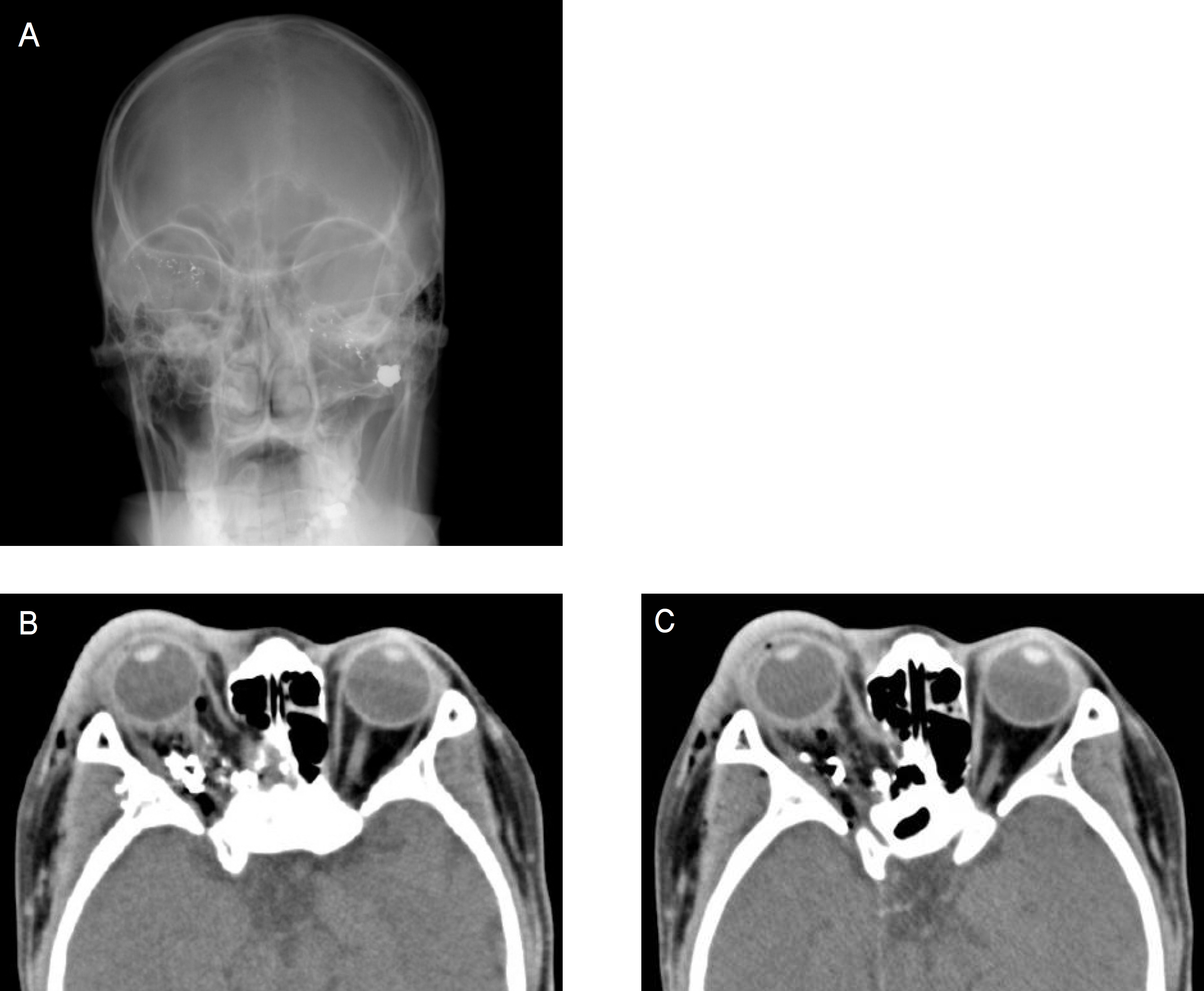
Figure 2. A large number of scattered metallic foreign bodies were detected from the left infratemporal fossa to the right temporal region (A). The metallic foreign bodies were detected in the right retrobulbar region, and an optic nerve injury was suspected. A fracture of the right medial orbital wall was detected, and a partial injury to the medial rectus muscle was suspected (B). A fracture of the right lateral wall was deteted, but no injury to the lateral rectus muscle was seen (C).

Figure 3. Mild limitation of adduction was noted 7 days after surgery (OD).
Cited by 1 articles
-
A Case of Intraorbital Foreign Body After Gunshot Wounds
Seong Taeck Kim, Dae Hyun Kim
J Korean Ophthalmol Soc. 2009;50(4):649-655. doi: 10.3341/jkos.2009.50.4.649.
Reference
-
References
1. Dinkel TA, Ward TP, Frey DM, Hollifield RD. Dissection along the optic nerve axis by a BB. Arch Ophthalmol. 1997; 115:673–5.
Article2. Dunya IM, Rubin PA, Shore JW. Penetrating orbital trauma. Int Ophthalmol Clin. 1995; 35:25–36.
Article3. Fuicher TP, McNab AA, Sullivan TJ. Clinical features and management of intraorbital foreign bodies. Ophthalmology. 2002; 109:494–500.
Article4. Karcioglu ZA, Nasr AM. Diagnosis and management of orbital inflammation and infections secondary to foreign bodies: a clinical review. Orbit. 1998; 17:247–69.
Article5. Turgut Y, Fatih SE, Huseyin Y, et al. Delayed trigeminocardiac reflex induced by an intraorbital foreign body. Opthalmologica. 2006; 220:65–8.6. Welti C, Mittleman R, Rao V. An atlas of forensic pathology. Chicago: American Society of Clinical Pathologies;1999.7. Jun YC, Lee SK, Kim SH, et al. Classification of the gun-shot wounds of the face by entrance and exit wounds of the bullet. J Korean Soc Plast Reconstr Surg. 1997; 24:1325–33.8. Sobaci G, Mutlu FM, Bayer A, et al. Deadly weapon-related open-globe injuries: outcome assessment by the ocular trauma classification system. Am J Ophthalmol. 2000; 129:47–53.
Article9. Kim MJ, Kim MH. Management of maxillo-facial gun-shot wound review of references and report of cases. J Korean Assoc Oral Maxillofac Surg. 1981; 7:51–60.10. Newman TL, Russo PA. Ocular sequelae of BB injuries to the eye and surrounding adnexa. J Am Optom Assoc. 1998; 69:583–90.11. Pieramici D, Sternberg P, Aaberg TM, et al. A system for classifying mechanical injuries of the eye (globe). Am J Ophthalmol. 1997; 123:820–31.
Article12. De Juan E, Sternberg P, Michels RS. Penetrating ocular injuries. Ophthalmology. 1983; 90:1318–22.
Article13. Esmaeli B, Elner SG, Schork A, Elner VM. Visual outcome and ocular survival after penetrating trauma: a clinicopathologic study. Ophthalmology. 1995; 102:393–400.14. Gilbert CM, Soong HK, Hirst LW. A two-year prospective study of penetrating ocular trauma at the Wilmer Ophthalmological Institute. Ann Ophthalmol. 1987; 19:104–6.15. Sternberg P, de Juan E, Michelle RG, Auer C. Multivariate analysis of prognostic factors in penetrating ocular injuries. Am J Ophthalmol. 1984; 98:467–72.
Article16. Shuttleworth GN, Galloway PH. Ocular air-gun injury: 19 cases. J R Soc Med. 2001; 94:396–9.
Article
- Full Text Links
-
- Actions
-
Cited
- CITED
-
- Close
- Share
-
- Similar articles
-
- Paraplegia Following Spinal Cord Contusion from an Indirect Gunshot Injury
- Assessment of Visual Outcome Following Optic Canal Decompression in Traumatic Optic Nerve Injuries
- Effect of High dose Corticosteroid and Optic Canal Decompression on Traumatic Optic Nerve Injury
- Traumatic Optic Neuropathy
- 2 Cases of Optic Nerve Decompression of Two Traumatic Optic Neuropathies Using Intranasal Endoscope
