J Korean Ophthalmol Soc.
2008 Jan;49(1):111-118. 10.3341/jkos.2008.49.1.111.
The Effect of Internal Limiting Membrane Peeling on Retinal Vein Occlusion Induced Macular Edema
- Affiliations
-
- 1Department of Ophthalmology, Kyungpook National University College of Medicine, Daegu, Korea. itkim@knu.ac.kr
- KMID: 2211190
- DOI: http://doi.org/10.3341/jkos.2008.49.1.111
Abstract
-
PURPOSE: To evaluate the effect of pars plana vitrectomy with indocyanine green-assisted peeling of the internal limiting membrane (ILM) on visual acuity in macular edema in RVO patients.
METHODS
Twenty-three eyes of 23 patients were treated consecutively (male: female=7:16). Thirteen patients were BRVO, and ten patients were CRVO. Vitrectomy that involved peeling the ILM with the assistance of indocyanine dye was performed in all 23 eyes. A visual acuity change of 2 lines or more was regarded as significant. We compared preoperative BCVA and postoperative BCVA.
RESULTS
Improvement of visual acuity was observed in 13 eyes (8 eyes in BRVO, 5 eyes in CRVO) of 23 total eyes (56.5%). No change in visual acuity was observed in 5 eyes (3 eyes in BRVO, 2 eyes in CRVO). Worsening of visual acuity was observed in 5 eyes (2 eyes in BRVO, 3 eyes in CRVO).
CONCLUSIONS
Pars plana vitrectomy with ILM peeling in patients with macular edema induced by RVO showed visual improvement in 56.5% of the cases in our study (61.5% in BRVO, 50% in CRVO). PPV with ILM peeling may be an effective procedure in reducing macular edema due to RVO.
MeSH Terms
Figure
-

Figure 1. Scatterplot illustrating visual acuity before and after ILM peeling in BRVO.
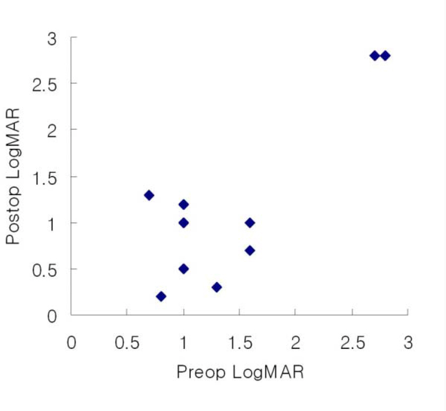
Figure 2. Scatterplot illustrating visual acuity before and after ILM peeling in CRVO.

Figure 3. Preoperative (A, B, C) and postoperative (D, E, F) fundus, fluorescein angiography and OCT appearance in case 13 with BRVO show reduced intraretinal hemorrhage (D), intraretinal leaking and stain (E), and macular thickness (F).
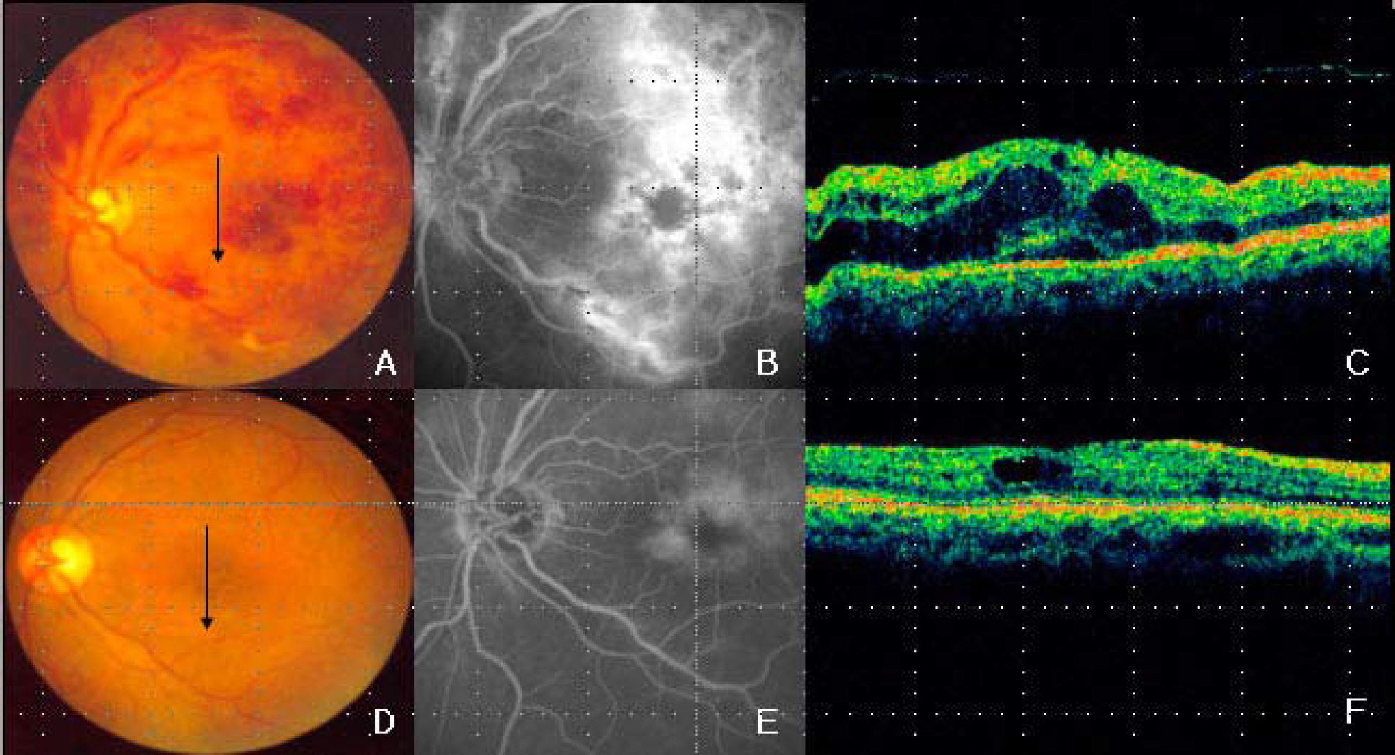
Figure 4. Preoperative (A, B, C) and postoperative (D, E, F) fundus, fluorescein angiography and OCT appearance in case 2 with CRVO show reduced intraretinal hemorrhage (D), intraretinal leaking and stain (E), and macular thickness (F).
Reference
-
References
1. Mandelcorn MS, Nrusimhadevara RK. Internal limiting membrane peeling for decompression of macular edema in retinal vein occlusion: A report of 14 cases. Retina. 2004; 24:348–55.
Article2. Radetzky S, Walter P, Fauser S, et al. Visual outcome of patients with macular edema after pars plana vitrectomy and indocyanine green-assisted peeling of the internal limiting membrane. Graefes Arch Clin Exp Ophthalmol. 2004; 242:273–8.
Article3. Eye Disease Case Control Study Group. Risk factors for branch retinal vein occlusion. Am J Ophthalmol. 1993; 116:286–96.4. Molnar I, Poitry S, Tsacopoulos M, et al. Effect of laser photocoagulation on oxygenation of the retina in miniature pigs. Invest Ophthalmol Vis Sci. 1985; 26:1410–4.5. Stefansson E, Machemer R, de Juan E Jr, et al. Retinal oxygenation and laser treatment in patients with diabetic retinopathy. Am J Ophthalmol. 1992; 113:36–8.6. The Central Vein Occlusion Study Group M report. Evaluation of grid pattern photocoagulation for macular edema in central vein occlusion. Ophthalmology. 1995; 102:1424–33.7. The Central Vein Occlusion Study Group. Natural History and Clinical Management of Central Retinal Vein Occlusion. Arch Ophthalmol. 1997; 115:486–91.8. Opremcak EM, Bruce RA, Lomeo MD, et al. Radial optic neurotomy for central retinal vein occlusion. A retrospective pilot study of 11 consecutive cases. Retina. 2001; 21:408–15.9. McAllister IL, Douglas JP, Constable IJ, Yu DY. Laser induced chorioretinal anastomosis for non ischemic central retinal vein occlusion: evaluation of the complications and their risk factor. Am J Ophthalmol. 1998; 126:219–29.10. Singh Hayreh S, Opremcak EM, Bruce RA, et al. Radial optic neurotomy for central retinal vein occlusion. Retina. 2002; 22:374–7.
Article11. Samuel MA, Desai UR, Gandolfo CB. Peripapillary retinal detachment after optic neurotomy for central vein occlusion. Retina. 2003; 23:580–3.12. Bresnick GH. Diabetic maculopathy: A critical review highlighting diffuse macular edema. Am J Ophthalmol. 1983; 1301–17.13. Porda SM, Krupin T, Asseff C, Beeker B. Topically administered steroid preparation. Arch Ophthalmol. 1971; 86:251.14. Opatowsky I, Feldman RM, Gross R, Feldman ST. Intraocular pressure elevation associated with inhalation and nasal corticosteroids. Ophthalmology. 1992; 102:177–9.
Article15. Lee J, Lee JH. The effect of intravitreal triamcinolone acetonide on cystoid macular edema. J Korean Ophthalmol Soc. 2004; 45:408–13.16. Kim IC, Ahn M, Cho NC. Intravitreal injection of triamcinolone acetonide for refractory diabetic macular edema. J Korean Ophthalmol Soc. 2004; 45:220–8.17. Sebag J, Balazs EA. Pathogenesis of cystoid macular edema: an anatomic consideration of vitreoretinal adhesion. Surv Ophthalmol. 1984; 28:S493–8.18. Avunduk AM, Centinkaya K, Kapicioglu Z, Kaya C. The effect of posterior vitreous detachment on the prognosis of branch retinal vein occlusion. Acta Ophthalmol Scand. 1997; 75:441–2.
Article19. Takahashi MK, Hikichi T, Akiba J, et al. Role of the vitreous and macular edema in branch retinal vein occlusion. Ophthalmic Surg Lasers. 1997; 28:294–9.20. Foos RY. Vitreoretinal juncture: topographical variations. Investigative Ophthalmology. 1972; 11:801–8.21. Hikichi T, Konno S, Trempe CL. Role of the vitreous in central retinal vein occlusion. Retina. 1995; 15:29–33.
Article22. Kado M, Jalkh AE, Yoshida A, et al. Vitreous change and macular edema in central retinal vein occlusion. Ophthalmic Surg. 1990; 21:544–9.23. Gandorfer A, Messmer EM, Ulbig MW, Kampik A. Resolution of diabetic macular edema after surgical removal of the posterior hyaloid and the inner limiting membrane. Am J Ophthalmol. 2000; 20:126–33.
Article24. Ikeda T, Sato K, Katano T, Hayashi Y. Improved visual acuity following pars plana vitrectomy for diabetic cystoid macular edema and detached posterior hyaloid. Retina. 2000; 20:220–2.
Article25. Terasaki H, Miyake Y, Nomura R, et al. Focal macular ERGs in eyes after removal of macular ILM during macular hole surgery. Invest Ophthalmol Vis Sci. 2001; 42:229–34.26. Haritoglou C, Gandorfer A, Gass CA, et al. The effect of indocyanine green on functional outcome of macular pucker surgery. Am J Ophthalmol. 2003; 135:328–37.27. Gandorfer A, Haritoglou C, Gass CA, et al. Indocyanine green-assisted peeling of the internal limiting membrane may cause retinal damage. Am J Ophthalmol. 2001; 132:431–3.
Article28. Maia M, Haller JA, Pieramici DJ, et al. Retinal pigment epithelial abnormalities after internal limiting membrane peeling guided by indocyanine green staining. Retina. 2004; 24:157–60.
Article29. Duff IF, Fall HF, Linman JW. Anticoagulant therapy in occlusive vascular disease of retina. Arch Ophthalmol. 1951; 46:601–17.30. Michel RG, Gass JD. The natural course of retinal branch vein obstruction. Trans Am Acad Ophthalmol Otolaryngol. 1974; 78:166–77.31. Lee DG, Cho NC. Combination of laser treatment and intravitreal triamcinolone injection for macular edema with branch retinal vein occlusion. J Korean Ophthalmol Soc. 2005; 46:287–96.32. Kim YG, Yu SY, Kwak HW. The effect of intravitreal triamcinolone acetonide injection according to the diabetic macular edema type. J Korean Ophthalmol Soc. 2005; 46:84–9.33. Kim YM, Kim SS, Lee SC, et al. Effects of pars plana vitrectomy comparing with intravitreal triamcinolone acetonide injection in diabetic macular edema. J Korean Ophthalmol Soc. 2006; 47:1072–9.
- Full Text Links
-
- Actions
-
Cited
- CITED
-
- Close
- Share
-
- Similar articles
-
- Arteriovenous Sheathotomy for Persistent Macular Edema in Branch Retinal Vein Occlusion
- Ultrastructure of the Internal Limiting Membrane Removed During Macular Hole and Diabetic Macular Edema Surgery
- Effect of Internal Limiting Membrane Peeling in Idiopathic Macular Holes Stage 3, 4
- Analysis of the Maular Change in Optical Coherent Tomography after Internal Limiting Membrane Peeling
- Eccentric Macular Hole Formation After Macular Hole Surgery
