J Korean Ophthalmol Soc.
2007 Oct;48(10):1410-1414. 10.3341/jkos.2007.48.10.1410.
A Case of Intraorbital Foreign Body Removed Using A Magnet Under C-arm Fluoroscopy
- Affiliations
-
- 1Department of Ophthalmology, College of Medicine Inje University, Pusan, Korea. eyeyang@inje.ac.kr
- KMID: 2210996
- DOI: http://doi.org/10.3341/jkos.2007.48.10.1410
Abstract
-
PURPOSE: We report a case of an intraorbital foreign body removed in a walk-in patient using a magnet under fluoroscopy.
METHODS
A patient walked into the eye clinic complaining of ocular pain caused by foreign body that pernetrated into his right lower eyelid while mowing the lawn one day before he came to the hospital. Since an orbital foreign body was observed when the patient entered the hospital, and a high-density metallic response was diagnosed within the orbit from a computerized tomogram, we performed an emergency operation to take out the foreign body within the orbit.
RESULTS
We removed the metallic foreign body, which was 5 mm in size and buried in the orbital fat, in an operation using a magnet under fluoroscopy.
CONCLUSIONS
This study shows that fluoroscopy and magnets are an efficient operative means of removing foreign bodies that are found within the orbital fat layer and are difficult to access.
Keyword
Figure
-
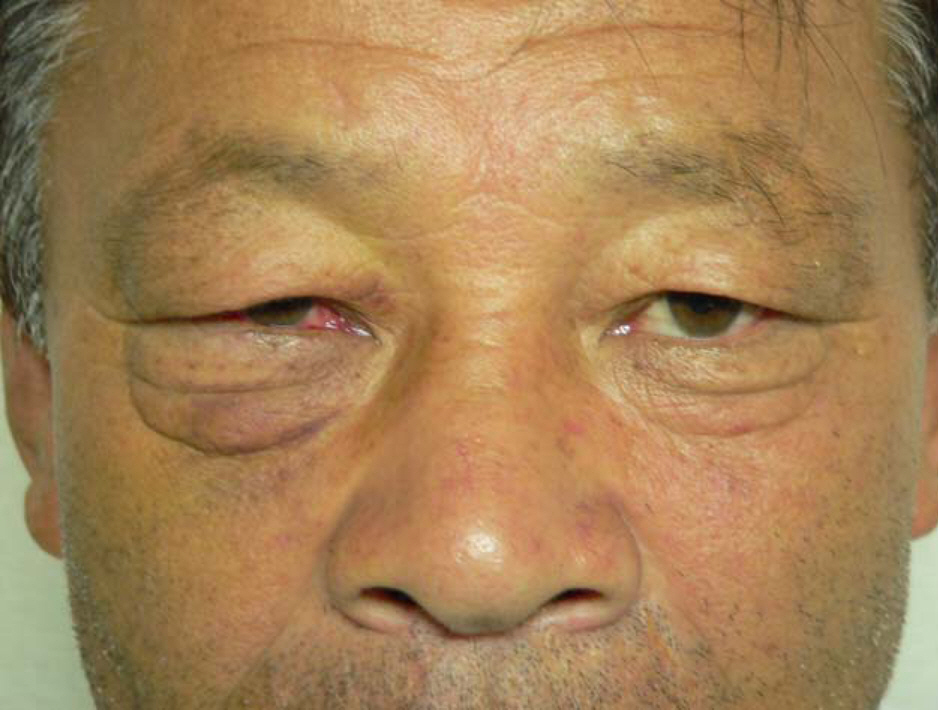
Figure 1. Preoparative photograph of the patient. The inlet (arrow) is not seen due to a skin fold and lower lid swelling. There is also some periorbital ecchymosis and conjunctival hemorrhage.
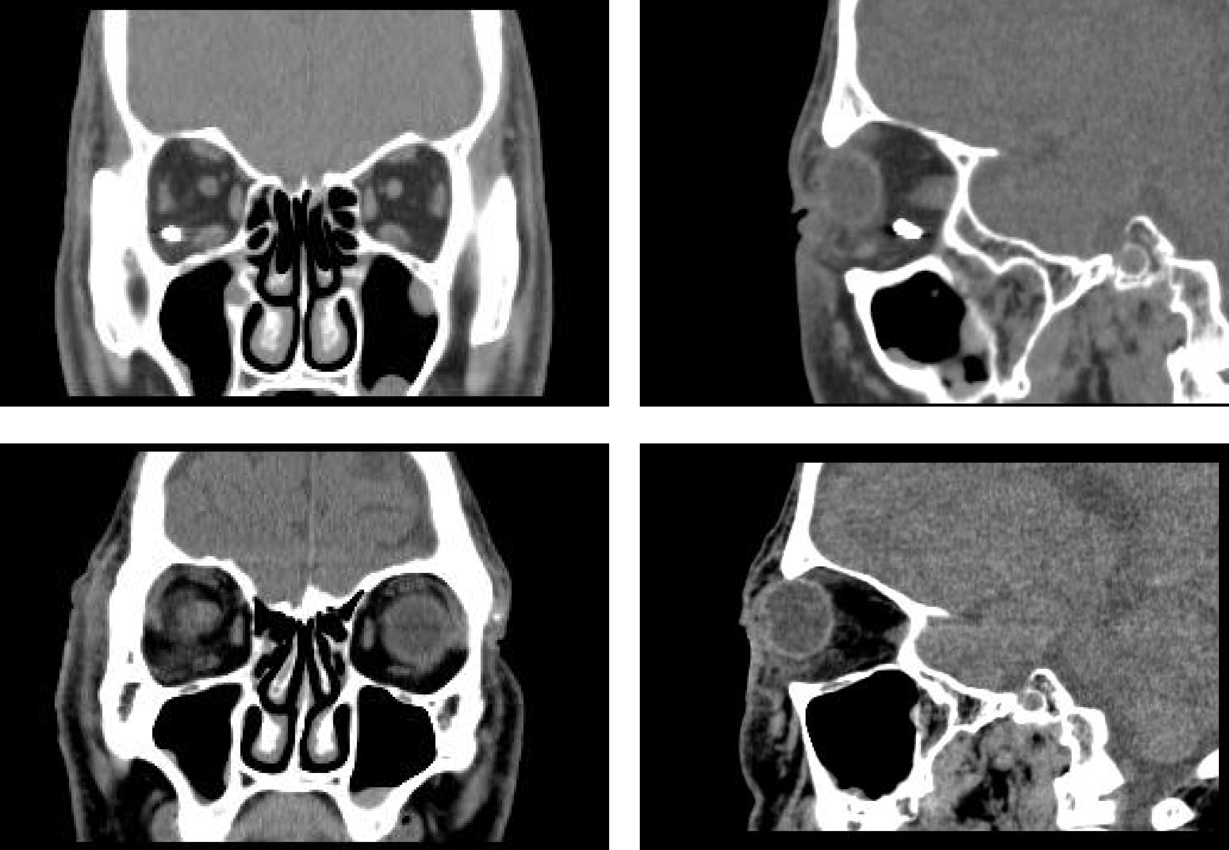
Figure 2. Preoperative (upper) and postoperative (lower) coronal and sagittal views of orbital CT scans. A foreign body with high density in the inferotemporal area of the orbit was removed by operation.
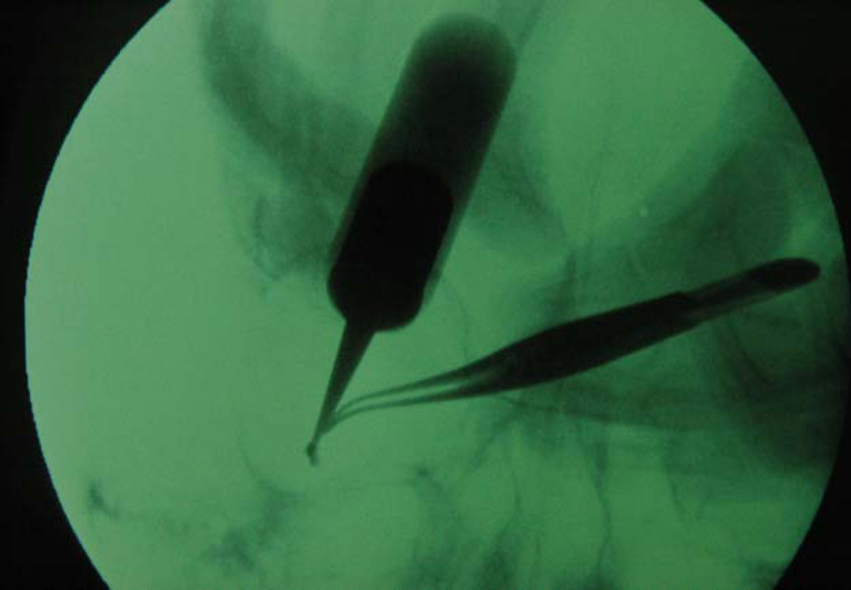
Figure 3. Intraoperative fluoroscopy with a magnet and forceps. Foreign body is captured by a magnet with assistance of forceps.
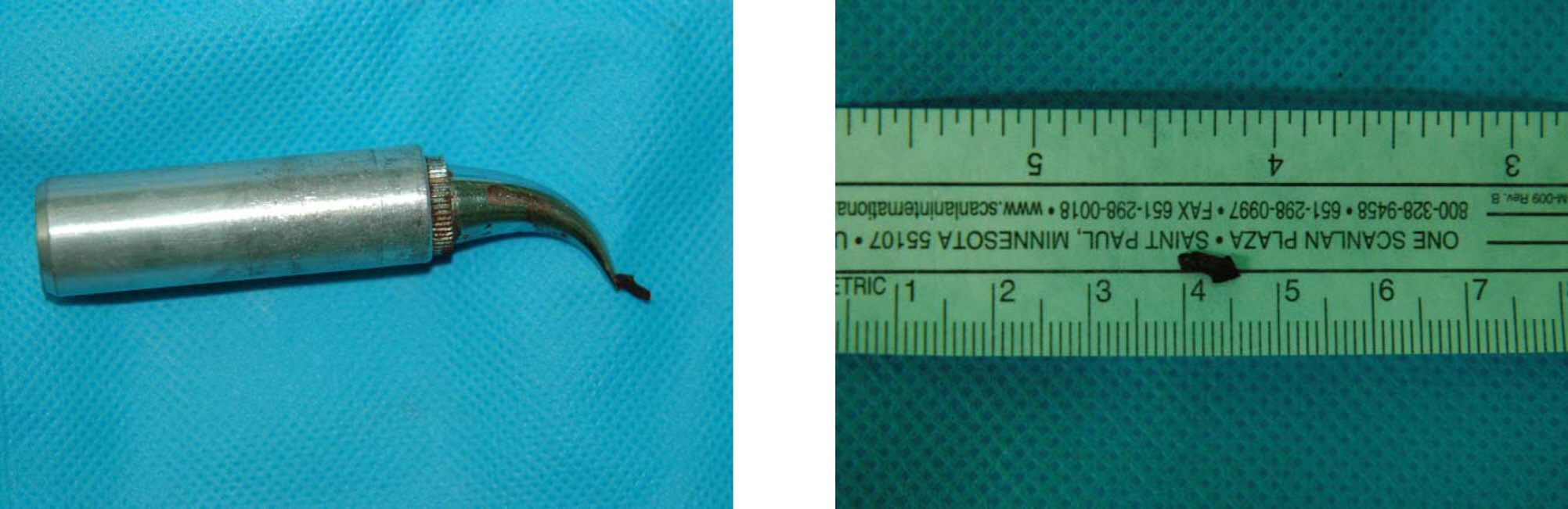
Figure 4. Removed foreign body on the tip of the magnet. The size of removed foreign body is about 5 mm in length and 2 mm in width.
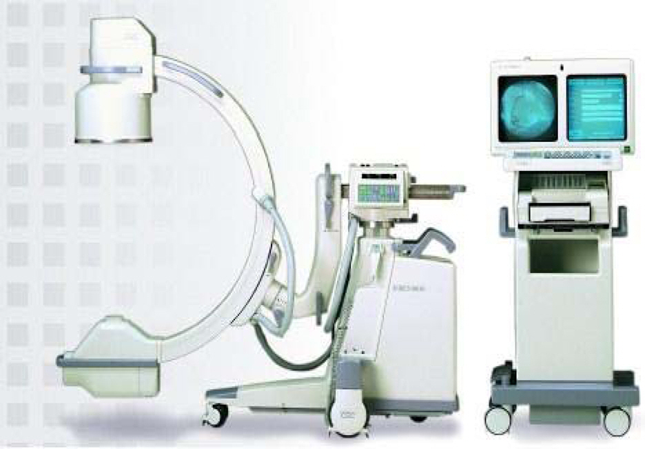
Figure 5. Digital Mobile Imaging System, OEC 9800 Plus, General Electric, USA.
Reference
-
References
1. Fulcher PF, McNab AA, Sullivan TJ. Clinical features and management of intraorbital foreign bodies. Ophthalmology. 2002; 109:494–500.
Article2. Ballen DH, Buckley ET. Removal of a large retrobulbar foreign body. Am J Ophthalmol. 1961; 52:715–7.
Article3. Park YG. A Case of Orbitocranial Foreign Body. J Korean Ophthalmol Soc. 1974; 26:386–9.4. Lim SJ, Lee JB, Hong YJ. A Case of Intraorbital Foreign Body. J Korean Ophthalmol Soc. 1985; 26:335–40.5. Macrae JA. Diagnosis and management of wooden orbital foreign body : case report. Br J Ophthalmol. 1979; 63:848–51.6. Michon JJ, Miller NR. Management of combined penetrating intraorbital and intracranial trauma. Arch Ophthalmol. 1993; 111:438–9.
Article7. Bartkowski SB, Kurek M, Stypalkowska J. Foreign bodies in the orbit, Review of 20 cases. J Maxillofac Surg. 1984; 12:97–102.8. Kim HK, Chung WS. Clinical Experience of Intraorbital Foreign Body. J Korean Ophthalmol Soc. 1997; 38:177–84.9. Charteris DG. Posterior penetrating injury of the orbit with retained foreign body. Br J Ophthalmol. 1988; 72:432–3.
Article10. Son OO, Kim CS, Lee JR. A Case of Intraorbital Foreign Body Diagnosed by Ultrasonography B - scan. J Korean Ophthalmol Soc. 1985; 26:547–9.11. Dallow RL. Diagnostic imaging in ophthalmology. 1st ed.New York: Springer-Verlag;1986. p. 55–69.12. Weisman RA. Computed Tomography in penentrating wounds of the orbit with retained foreign body. Arch Otolaryngol. 1983; 109:265–8.13. Etherington RJ. Localization of intraocular and intraorbital foreign bodies using CT. Clin Radiol. 1989; 40:610–4.14. Rahman NU, Jamjoom A, Jamjoom ZA. Orbito-cranial injury caused by penetrating metallic foreign bodies: report of two cases. Int Ophthalmol. 1997; 21:13–7.15. Spoor TC, Nesi FA. Management of ocular, orbital and adnexal trauma. 1st ed.New York: Raven press;1988. p. 271–92.16. Duke-Elder S, MacFaul PA. Injuries - part 1, Mechanical Injuries. In : Duke-Elder S, editor. System of Ophthalmology. London: Henry Kimpton;1972. v. 14. chap. 1.17. Duke-Elder S, MacFaul PA. System of Ophthalmology. 1st ed.14. London: Henry Kimpton;1972. p. 655–69.18. Rootman J, Neigel JM. Diseases of the Orbit. 2nd ed.Philadelphia: Lippincott;1988. p. 504–20.19. Tse DT. Color atlas of opthalmic surgery: Oculaoplastic surgery. 1st ed.12. Philadelphia: J.B. Lippincott;1992. p. 445–62.20. Merriam GR, Focht EF. Clinical study of radiation cataracts and the relationship to dose. Amer J Roentgenol. 1957; 77:759–85.21. Merriam GR, Worgul BV. Experimental radiation cataract: its clinical relevance. Bull N Y Acad Med. 1983; 59:372–92.22. Britten MJA, Halnan KE, Meredith WJ. Radiation cataract: new evidence on radiation dosage to the lens. Br J Radiol. 1966; 39:612–7.
- Full Text Links
-
- Actions
-
Cited
- CITED
-
- Close
- Share
-
- Similar articles
-
- Fluroscopic Removal of the Foreign Bodies from Gastroesophagus Using the Magnet
- Case Report of Retained Intraorbital Metallic Foreign Body Removal
- Successful Localization of Intraoral Foreign Body with C-arm Fluoroscopy
- Retrieval of Metallic Foreign Bodies from the Upper Airway Using Intraoperative C-Arm Fluoroscopy: Case Report and Literature Review
- Two Cases of Intraorbital Wooden Foreign Bodies
