J Korean Soc Spine Surg.
2008 Dec;15(4):243-249. 10.4184/jkss.2008.15.4.243.
Metal Failure in patients with Short-segmental Pedicle Screw Fixation and Fusion for Degenerative Lumbar Disease: Comparison Monoaxial with Polyaxial Screw
- Affiliations
-
- 1Department of Orthopaedic Surgery, Eulji University School of Medicine, Daejeon, Korea. jwkang@eulji.ac.kr
- 2Department of Orthopaedic Surgery, Daejeon Veterans Hospital, Daejeon, Korea.
- 3Department of Orthopaedic Surgery, Hongseong Medical Center, Hongseong, Korea.
- KMID: 2209627
- DOI: http://doi.org/10.4184/jkss.2008.15.4.243
Abstract
- STUDY DESIGN: Retrospective study
OBJECTIVE
To evaluate the factors affecting metal failure and screw loosening of short-segmental (1- or 2-segmental) monoaxial or polyaxial screw fixation for degenerative lumbar disease. SUMMARY OF LITERATURE REVIEW: There was a report on metal failure and screw loosening in short-segmental monoaxial and polyaxial screw fixation in degenerative lumbar disease.
MATERIALS AND METHODS
This study examined 227 cases who underwent short-segmental transpedicular screw fixation and vertebral fusion for a degenerative lumbar.
RESULTS
Metal failure of transpedicular screws was detected in 6 cases, 3 each in groups A and B. Screw loosening occurred in 16 and 43 cases in group A and B, respectively. Both groups had a similar incidence of spinal stenosis with instability and spondylolisthesis. The failure rate and screw loosening according to the fusion level was also similar. The failure and screw loosening rates was higher in the cases who did not undergo PLIF than in the cases who underwent PLIF but the difference was not statistically significant.
CONCLUSION
The metal failure and screw loosening rates after transpedicular screw fixation and spinal fusion procedures for degenerative lumbar diseases using monoaxial screws and polyaxial screws were similar.
Keyword
Figure
-
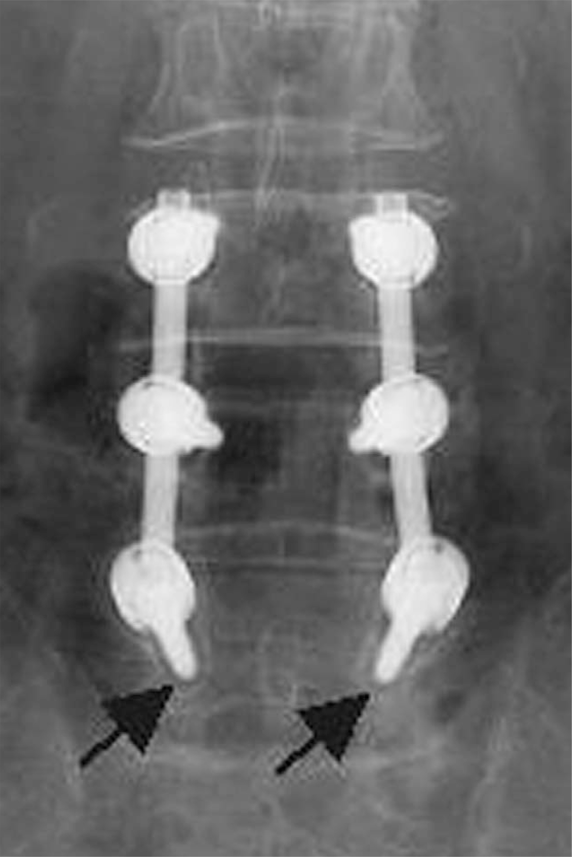
Fig. 1. 59-year-old patient with spinal stenosis of the lumbar spine. A continuous lucency at the screw-bone interface surrounded by a thin radiolucent zone (halo sign-black arrows) on the AP radiograph indicates loosening of both distal screws on 4 months follow up.
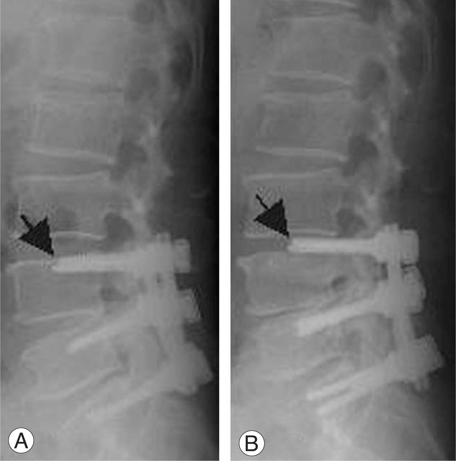
Fig. 2. 62-year-old patient with spinal stenosis shows pull-out (black arrow) of screws from the vertebral body due to increase of kyphosis of the lumbar spine on 4 months (A) and 10 months (B) follow up.
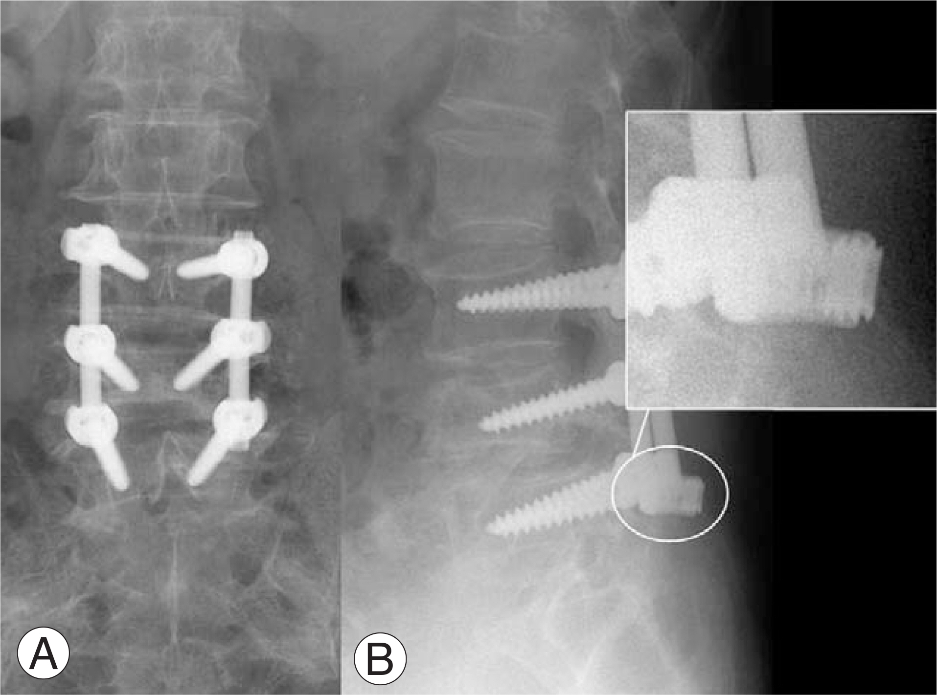
Fig. 3. 67-year-old patient with spinal stenosis shows screw-rod dissociation on 2 months follow up.
Reference
-
01). Sanden B., Olerud C., Petren-Mallmin M., Johansson ., Larsson S. The significance of radiolucent zones surrounding pedicle screws: Definition of screw loosening in spinal instrumentation. J Bone and Joint Surg Br,. 2004. 86:457–461.02). Krag MH., Beynnon BD., Pope MH., Frymoyer JW., Haugh LD. An internal fixation for posterior application to short segments for the thoracic, lumbar, or lumbosacral spine: Design and testing. Clin. Orthop. 1986. 203:75–98.03). Roy-Camille R., Saillant G., Mazel C. Plating or thoracic, thoracolumbar and lumbar injuries with pedicle screw plates. Ortho. Clin. North Am,. 1986. 17:147–159.04). Boucher HH. A method of spinal fusion. J Bone and Joint Surg Br,. 1959. 41:248–259.
Article05). Harrington PR., Tullos HS. Reconstruction of severe spondylolisthesis in children. South Med J,. 1969. 62:1–7.06). Steffee A., Biscup R., Stickowski D. Segmental spine plates with pedicle screws fixation: A new internal fixation device for disorders of the lumbar and thoracic spine. Clin. Orthop. 1986. 203:45–53.07). Lee KY., Kim CH., Song CG. Metal failure of pedicle screw system. J Kor Soc Surg,. 2002. 9:157–163.
Article08). Shin BJ., Kim KJ., Kim ST., Kim YI. Survivorship analysis of pedicle screw fixation. J Kor Soc Surg,. 1999. 6:355–361.09). McAfee PC., Farey ID., Sutterlin CE., Gurr KR., Wrden KE., Cunningham BW. The effect of spinal implant rigidity on vertebral bone density: a canine model. Spine,. 1991. 16:190–197.10). Okuyama K., Abe K., Suzuki T., Tamura Y., Chiba M., Sato K. Posterior lumbar interbody fusion. A retrospective study of complication after facet joint excision and pedicle screw fixation in 148 cases. Acta Orthop Scand,. 1999. 10:329–334.11). Ohlin A., Karlsson M., Dupe H., Harserius R., Redlund-Hohnell I. Complication after transpedicular stabilization of the spine. Spine. 1994. 19:2774–2779.12). Schatzker J., Horne JG., Sumner-Smith G. The effect of movement on the holding power of screws in bone. Clin. Orthop. 1975. 111:257–262.13). Kuklo TR., Potter BK., Polly DW., Lenke LG. Monaxial versus thoracic pedicle screws in the correction of adolescent idiopathic scoliosis. Spine,. 2005. 30:2113–2120.14). Stanford RE., Loefler AH., Stanford PM., Walsh WR. Multiaxial pedicle screw designs: Static and dynamic mechanical testing. Spine,. 2004. 29:367–375.
Article15). Fogel GR., Reitman CA., Liu W., Esses SI. Physical characteristics of polyaxial-head pedicle screws and biomechanical comparison of load with their failure. Spine,. 2003. 28:470–473.16). Chen SH., Lin RM., Chen HH., Tsai KJ. Biomechanical effects of ployaxial pedicle screw fixation on the lumbosacral segments with an anterior interbody cage support. BMC Musculoskeletal Disorder,. 2007. 8:28.
Article
- Full Text Links
-
- Actions
-
Cited
- CITED
-
- Close
- Share
-
- Similar articles
-
- An Innovative Universal Screw Removal Instrument
- Unilateral versus Bilateral Pedide Scrwe Fixation in Lumbar Spinal Fusion
- Comparison between Posterolateral Fusion with Pedicle Screw Fixation and Anterior Interbody Fusion with Pedicle Screw Fixation in Spondylolytic Spondylolisthesis of the Lumbar Spine
- Posterior Atalntoaxial Fusion with C1 Lateral Mass Screw and C2 Pedicle Screw Supplemented with Miniplate Fixation for Interlaminar Fusion : A Preliminary Report
- The Comparison of Changes in the Dimensions of the Intervertebral Disc and Neural Foramen between Anterior Lumbar Interbody Fusion and Posterolateral Fusion in the Lumbar Spine
