Inverted Meckel Diverticulum in the Jejunum with CT Demonstration of Vitelline Artery and Veins: A Case Report
- Affiliations
-
- 1Department of Radiology, Dongsan Medical Center, Keimyung University School of Medicine, Korea. kjh2603@dsmc.or.kr
- 2Department of Pathology, Dongsan Medical Center, Keimyung University School of Medicine, Korea.
- 3Department of Radiology, Dongrae Paik Hospital, Inje University College of Medicine, Korea.
- KMID: 2208996
- DOI: http://doi.org/10.3348/jksr.2010.62.1.61
Abstract
- Meckel diverticulum arising in the jejunum is a very rare condition, and inverted Meckel diverticulum of the jejunum has not been described previously. We report a case of inverted Meckel diverticulum of the jejunum with the computed tomography (CT) observations of the vitelline artery and veins in a 28-year-old woman. CT demonstrated an elongated intraluminal mass with a central area of fat attenuation in the jejunum as well as a vitelline artery and veins in the central area of this mass.
Figure
-
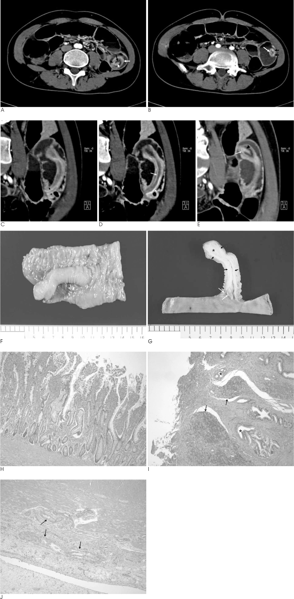
Fig. 1 A 28-year-old woman with an inverted Meckel diverticulum in the distal jejunum. A. Axial contrast-enhanced abdominal CT scan in the arterial phase shows an oval ring-shaped mass within the dilated small bowel of the left mid abdomen. This mass represents the tip of an inverted Meckel diverticulum and has an alternating outer diverticular wall (white long arrow), inner mesenteric fat (long black arrow), and a central ectopic pancreatic tissue (short white arrow). A vitelline artery can be seen (short black arrow). B. Axial contrast-enhanced abdominal CT scan in the arterial phase 2 cm below (A) shows a ring-shaped mass with a single vetelline artery (long white arrow) and two vitelline veins (short black arrows). C-E. Coronal-oblique reconstruction images (C-E, from anterior to posterior) along the long axis of the inverted diverticulum show an elongated, smoothly marginated intraluminal mass parallel to the long axis of the bowel with a central area of fat attenuation surrounded by a thick collar of soft-tissue attenuation. Vitelline artery (long arrows), vitelline veins (short arrows), and ectopic pancreatic tissue (arrowheads) are well visualized. F. The gross pathologic specimen shows an elongated, longitudinal diverticular mass protruding from the pale pink to tan mucosa of distal jejunum, measuring 6.5 cm in length and 2.0 cm in diameter. The ruler indicates centimeters. G. A cut section of the diverticular mass revealed adipose tissue (asterisk) in the tip sandwiched between the continuous lining muscularis layers (short arrows). A few vitelline vessels of the diverticular root (long arrows) are running along the inner serosal wall of the diverticular mass. The ruler indicates centimeters. H. The mucosa of an inverted diverticular mass in the distal jejunum shows relatively tall slender villi and dense infiltration of inflammatory cells (H & E, ×200). I. The tip of the inverted diverticular mass shows focal erosive mucosa and underlying granulation tissue, as well as pancreatic ducts (asterisks) and acinar structures (arrows) (H & E, ×200). J. The inner serosal wall of the inverted diverticulum shows vitelline vessels (arrows) in the extramuscular dense fibroadipose tissue (H & E, ×200).
Reference
-
1. Pantongrag-Brown L, Levine MS, Elsayed AM, Buetow PC, Agrons GA, Buck JL. Inverted Meckel diverticulum: clinical, radiologic, and pathologic findings. Radiology. 1996; 199:693–696.2. Dujardin M, de Beeck BO, Osteaux M. Inverted Meckel's diverticulum as a leading point for ileoileal intussusception in an adult: case report. Abdom Imaging. 2002; 27:563–565.3. Shindoh N, Kurosaki A, Ozaki Y, Kyogoku S, Sumi Y, Katayama H. Characteristic angiographic appearance of inverted Meckel's diverticulum. AJR Am J Roentgenol. 1997; 169:1569–1571.4. Mitchell AW, Spencer J, Allison DJ, Jackson JE. Meckel's diverticulum: angiographic findings in 16 patients. AJR Am J Roentgenol. 1998; 170:1329–1333.5. Kouraklis G, Glinavou A, Mantas D, Kouskos E, Karatzas G. Clinical implications of small bowel diverticula. Isr Med Assoc J. 2002; 4:431–433.6. Yovich JV, Horney FD. Congenital jejunal diverticulum in a foal. J Am Vet Med Assoc. 1983; 183:1092.7. McSwain GR, Anderson MC. Meckel's diverticulum of the proximal jejunum. Arch Surg. 1979; 114:212–213.8. Miyoshi S, Ikeda M, Kido T, Matsuda Y, Fukada R, Nakajima K, et al. Abnormal persistence of the right vitelline vein. J Pediatr Surg. 1984; 19:204–205.9. Monedero MD, Ripolles T, Nicolau MJ, Martinez-Perez MJ. Pancreatic pseudotumor in Meckel diverticulum. Abdom Imaging. 2006; 31:688–690.10. Hollerweger A, Rieger S, Hübner E, Macheiner P. Sonographic diagnosis of an inverted Meckel diverticulum. J Ultrasound Med. 2007; 26:1263–1266.
- Full Text Links
-
- Actions
-
Cited
- CITED
-
- Close
- Share
-
- Similar articles
-
- A Clinical Study of Vitelline Duct and Vessel Remnants
- Intussusception due to Inverted Meckel Diverticulum with Ectopic Pancreas: A Case Report
- A Case of Massive Hematochezia from a Meckel's Diverticulum without Ectopic Mucosa
- Axial Torsion of Meckel's Diverticulum Causing Small Bowel Obstruction in Adult: A Case Report
- Intussusception in an Adult due to Inverted Meckel's Diverticulum with Ectopic Pancreatic Tissue
