Variation of Internal Mammary Vessels: Vein Lateral to Artery: Two Cases
- Affiliations
-
- 1Department of Radiology, Seoul St. Mary's Hospital, The Catholic University of Korea College of Medicine, Seoul, Korea. eclipse831211@gmail.com
- KMID: 2208817
- DOI: http://doi.org/10.3348/jksr.2013.69.2.139
Abstract
- Congenital variations of the internal mammary vascular anatomy are rare and may be encountered during various percutaneous transthoracic procedures. Hereby, we present two cases of congenital variations of the internal mammary vessel incidentally found on CT, with the vein entirely or partially lying lateral to the artery in the first intercostals space. The imaging findings and the clinical implications of these rare anatomical variations are being presented.
Figure
-

Fig. 1 A 25-year-old female with a palpable supraclavicular mass. A. Coronal maximum-intensity-projection CT image of a 25-year-old female (Case 1). The right (arrowheads) and left internal mammary arteries run laterally to their concomitant veins (arrows) in the first intercostals space, above which the vein cross over the arteries to join the brachiocephalic veins (B). B. CT scan shows a biopsy needle (arrowhead) advanced lateral to internal mammary vessels (arrow) into an anterior mediastinal mass, which was subsequently confirmed as diffuse large B cell lymphoma. Note.-I = innominate artery

Fig. 2 Coronal maximum-intensity-projection CT image of a 73-year-old male with a lung mass (Case 2). The right internal mammary artery (IMA; white arrow) is running medially to the right internal mammary vein (IMV; arrowhead) in the first intercostal space. The left IMV (black arrow) crosses over the left IMA (vein anterior to artery on axial plane) in the middle of the first intercostal space.
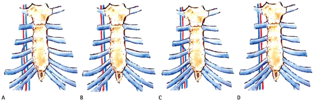
Fig. 3 Four patterns of IMV anatomy. Modified from Arnez ZM, Valdatta L, Tyler MP, Planinsek F. Br J Plast Surg 1995;48:540-545 (1). Blue: internal mammary vein. Red: internal mammary artery. (A) Type 1. (B) Type 2. (C) Type 3. (D) Type 4. Type 1 and 3: a single IMV lying medial (lateral) to the IMA dividing into two tributaries usually at the level of the 3rd or 4th intercostal space. Type 2 and 4: a single IMV runs medially (laterally) to the artery throughout its course, with no divisions. Note.-IMA = internal mammary artery, IMV = internal mammary vein
Reference
-
1. Arnez ZM, Valdatta L, Tyler MP, Planinsek F. Anatomy of the internal mammary veins and their use in free TRAM flap breast reconstruction. Br J Plast Surg. 1995; 48:540–545.2. Glassberg RM, Sussman SK. Life-threatening hemorrhage due to percutaneous transthoracic intervention: importance of the internal mammary artery. AJR Am J Roentgenol. 1990; 154:47–49.3. Glassberg RM, Sussman SK, Glickstein MF. CT anatomy of the internal mammary vessels: importance in planning percutaneous transthoracic procedures. AJR Am J Roentgenol. 1990; 155:397–400.4. Han S, Yoon SY, Park JM. The anatomical evaluation of internal mammary vessels using sonography and 2-dimensional computed tomography in Asians. Br J Plast Surg. 2003; 56:684–688.5. Mazeh H, Alaiyan B, Vald O, Mizrahi I, Klimov A, Eid A, et al. Internal mammary artery injury during central venous catheter insertion for TPN: rare but fatal. Nutrition. 2010; 26:849–851.6. Jansen EW, Lampmann LE, Lohle PN, van Rooy WJ, Pasteuning WH. False aneurysm of the right internal mammary artery. Vasa. 1999; 28:213–214.7. Kruyt PM, Winter LH, Koning J. A pseudo-aneurysm of the internal mammary artery: a very rare complication of subclavian vein puncture. Eur J Vasc Surg. 1993; 7:349–351.8. Gupta S, Seaberg K, Wallace MJ, Madoff DC, Morello FA Jr, Ahrar K, et al. Imaging-guided percutaneous biopsy of mediastinal lesions: different approaches and anatomic considerations. Radiographics. 2005; 25:763–786. discussion 786-788.9. Westcott JL. Percutaneous needle aspiration of hilar and mediastinal masses. Radiology. 1981; 141:323–329.
- Full Text Links
-
- Actions
-
Cited
- CITED
-
- Close
- Share
-
- Similar articles
-
- The Anatomical Study of Internal Mammary Perforators
- Preparation of the internal mammary artery graft in coronary artery surgery-comparison of free mammary artery flows
- The Anatomical Study of Internal Mammary Vessels Using Sonography and 2-Dimensional Computed Tomography
- Comparison of the second and third intercostal spaces regarding the use of internal mammary vessels as recipient vessels in DIEP flap breast reconstruction: An anatomical and clinical study
- Selection of Recipient Vessels in Delayed Breast Reconstruction with Free TRAM Flap
