Cervical Spinal Monostotic Fibrous Dysplasia: A Case Report
- Affiliations
-
- 1Department of Radiology, Haeundae Paik Hospital, Inje University College of Medicine, Busan, Korea. okkimmd@hanafos.com
- KMID: 2208807
- DOI: http://doi.org/10.3348/jksr.2013.69.3.239
Abstract
- Monostotic fibrous dysplasia of the cervical vertebra is quite unusual. The author reports a case of monostotic fibrous dysplasia affecting the second cervical vertebra with descriptions from the CT, MR and bone scanning findings.
Figure
-
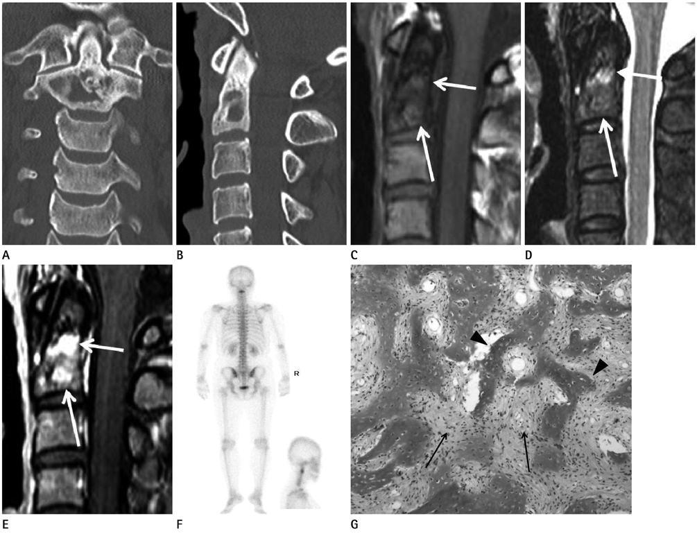
Fig. 1 Monostotic fibrous dysplasia of the C2 in a 42-year-old woman. A, B. Coronal (A) and sagittal (B) CT scan of C-spine reveal the irregular margined osteolytic lesion with peripheral bony sclerosis, without evidence of the bony cortical destructions, in C2 body. C-E. Sagittal MRI reveal the irregular margined osseous lesion incompletely surrounded by the rim of low signal intensity in the C2 body (arrows in C, D), showing low to intermediate SI on T1WI (C), heterogeneous mixed, low to high, SI on STIR image (D), and marked contrast enhancements (arrows in E) on Gd-T1WI (E) at the sites corresponding to the osteolytic areas of CT scan. F. Bone scan following administration of Tc-99m MDP reveal the focal tracer uptake in the upper cervical spine. Note no other skeletal tracer uptake in the whole body scan. G. Photomicrograph of surgical specimen shows classic microscopic appearance of fibrous dysplasia consisting of irregular immature trabeculae of woven bone with lack of osteoblastic rimming (arrowheads) in bland fibroblastic and collagenous matrix (arrows) (H&E stain, × 40). Note.-Gd = gadolineum, MDP = methylene diphosphonate, SI = signal intensity, STIR = short tau inversion recovery, T1WI = T1-weighted image
Reference
-
1. Arantes M, Vaz AR, Honavar M, Resende M, Pereira JR. Fibrous dysplasia of the first cervical vertebra. Spine (Phila Pa 1976). 2008; 33:E933–E935.2. Proschek D, Orler R, Stauffer E, Heini P. Monostotic fibrous dysplasia of the spine: report of a case involving a cervical vertebra. Arch Orthop Trauma Surg. 2007; 127:75–79.3. Schoenfeld AJ, Koplin SA, Garcia R, Hornicek FJ, Mankin HJ, Raskin KA, et al. Monostotic fibrous dysplasia of the spine: a report of seven cases. J Bone Joint Surg Am. 2010; 92:984–988.4. Gogia N, Marwaha V, Atri S, Gulati M, Gupta R. Fibrous dysplasia localized to spine: a diagnostic dilemma. Skeletal Radiol. 2007; 36:Suppl 1. S19–S23.5. Mauras N, Blizzard RM. The McCune-Albright syndrome. Acta Endocrinol Suppl (Copenh). 1986; 279:207–217.6. Dreizin D, Glen C, Jose J. Mazabraud syndrome. Am J Orthop (Belle Mead NJ). 2012; 41:332–335.7. Kransdorf MJ, Moser RP Jr, Gilkey FW. Fibrous dysplasia. Radiographics. 1990; 10:519–537.8. Park SK, Lee IS, Choi JY, Cho KH, Suh KJ, Lee JW, et al. CT and MRI of fibrous dysplasia of the spine. Br J Radiol. 2012; 85:996–1001.9. Asazuma T, Sato M, Masuoka K, Yasuoka H, Tsuji T, Aida S. Monostotic fibrous dysplasia of the lumbar spine: case report and review of the literature. J Spinal Disord Tech. 2005; 18:535–538.
- Full Text Links
-
- Actions
-
Cited
- CITED
-
- Close
- Share
-
- Similar articles
-
- Monostotic Fibrous Dysplasia of the Cervical Spine: A Case Report
- Pathologic Fracture in Cervical Spine with Monostotic Fibrous Dysplasia: Case Report
- Monostotic Fibrous Dysplasia in the Metacarpal Bone: A Case Report
- Osteosarcoma Arising in Monostotic Fibrous Dysplasia of the Femur: A Case Report
- Monostotic Fibrous Dysplasia of Inferior Turbinate
