A Case Report of Congenitally Corrected Transposition of Great Arteries: Morphologic and Functional Evaluation with Cardiac CT
- Affiliations
-
- 1Department of Radiology, College of Medicine, Soonchunhyang University, Bucheon Hospital, Bucheon, Korea. acarad@naver.com
- 2Department of Cardiology, College of Medicine, Soonchunhyang University, Seoul Hospital, Seoul, Korea.
- KMID: 2208801
- DOI: http://doi.org/10.3348/jksr.2013.69.3.197
Abstract
- Congenitally corrected transposition of the great arteries (ccTGA) is a rare congenital anomaly characterized by atrioventricular and ventriculoarterial discordance. We report a case of new-onset heart failure in a 69-year-old female in whom cardiac CT demonstrated ccTGA without the associated cardiovascular anomalies. In this case, cardiac CT was useful for elucidating the rare and unexpected congenital etiologies of abrupt-onset heart failure in an old patient by the simultaneous evaluation of cardiac morphology and function as a single study (inserted).
MeSH Terms
Figure
-
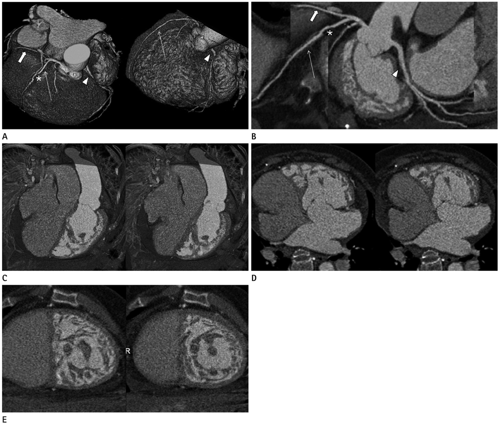
Fig. 1 Cardiac CT performed on 69-year-old female. A. Three-dimensional volume-rendered images show spatial relationship of great arteries with ascending aorta and main pulmonary artery. Anterior descending artery (long arrow) and circumflex artery (short arrow) arise from left main coronary artery off of anterior aortic sinus. Note stair-step artifact (*) from irregular heart rhythm on mid segment of left anterior descending artery. Right coronary artery (arrowhead) originates from posterior aortic sinus. B. Curved multiplanar reformation image demonstrates anterior descending (long arrow), circumflex (short arrow), and right coronary artery (arrowhead) without luminal stenosis. Again, stair-step artifact (*) is noted on mid segment of left anterior descending artery. C, D. Diastolic (left) and systolic (right) reconstruction images clearly depict systemlic right ventricle and morphologic left ventricle on 3 dimensional volume rendering image (C) and 4 chamber view (D). E. Diastolic (left) and systolic (right) short axis reconstructions show hypertrabeculation and intertrabecular recesses filled with blood.
Reference
-
1. Wallis GA, Debich-Spicer D, Anderson RH. Congenitally corrected transposition. Orphanet J Rare Dis. 2011; 6:22.2. Kharge J, Prasad MR, Ramegowda RT. An unusual case of congenitally corrected transposition of the great arteries associated with noncompaction-like remodeling of the morphological right ventricle. Echocardiography. 2011; 28:E212–E214.3. Nagle JP, Cheitlin MD, McCarty RJ. Corrected transposition of the great vessels without associated anomalies: report of a case with congestive failure at age 45. Chest. 1971; 60:367–370.4. Graham TP Jr, Bernard YD, Mellen BG, Celermajer D, Baumgartner H, Cetta F, et al. Long-term outcome in congenitally corrected transposition of the great arteries: a multi-institutional study. J Am Coll Cardiol. 2000; 36:255–261.5. Patrignani A, D'Aroma A, Cicogna S. Unusual association between "congenitally corrected transposition of the great arteries" and "noncompaction" of the right systemic ventricle. Int J Cardiovasc Imaging. 2009; 25:551–553.6. Purvis J, Barr S. An appearance of "non-compaction" of the right systemic ventricle is common in "congenitally corrected transposition of the great arteries. Int J Cardiovasc Imaging. 2009; 25:751–752.7. Ruzsics B. Integrative computed tomography imaging of ischemic heart disease. J Thorac Imaging. 2010; 25:231–238.8. Chang DS, Barack BM, Lee MH, Lee HY. Congenitally corrected transposition of the great arteries: imaging with 16-MDCT. AJR Am J Roentgenol. 2007; 188:W428–W430.
- Full Text Links
-
- Actions
-
Cited
- CITED
-
- Close
- Share
-
- Similar articles
-
- Transposition of the Great Arteries: Historical Background
- An Adult Case of Congenitally Corrected Transposition of the Great Arteries Associated with Paroxysmal Atrial Fibrillation and Heart Failure
- Congenitally Corrected Transposition of the Great Arteries
- Right Ventricular Inflow Obstruction Caused by Supratricuspid Ring after the Conventional Biventricular Repair of Congenitally Corrected Transposition of Great Arteries: A case report
- A case of isolated congenitally corrected transposition of the great arteries with complete atrioventricular block
