J Korean Soc Radiol.
2014 Nov;71(5):258-264. 10.3348/jksr.2014.71.5.258.
Relationships between Rotator Cuff Tear Types and Radiographic Abnormalities
- Affiliations
-
- 1Department of Diagnostic Radiology, College of Medicine, Chungbuk National University, Cheongju, Korea. ka1000@cbnuh.or.kr
- KMID: 2208796
- DOI: http://doi.org/10.3348/jksr.2014.71.5.258
Abstract
- PURPOSE
To determine relationships between different types of rotator cuff tears and radiographic abnormalities.
MATERIALS AND METHODS
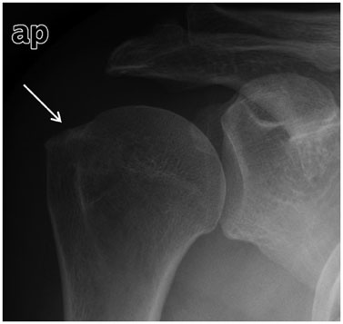
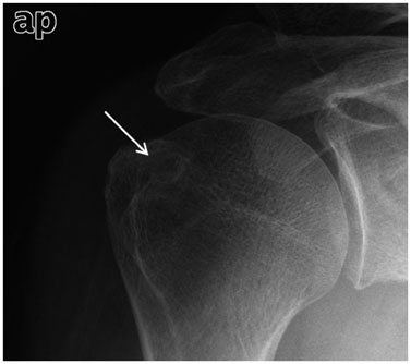
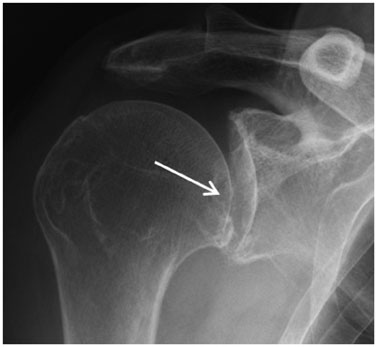
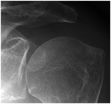
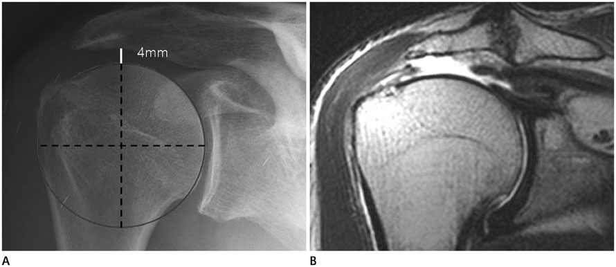
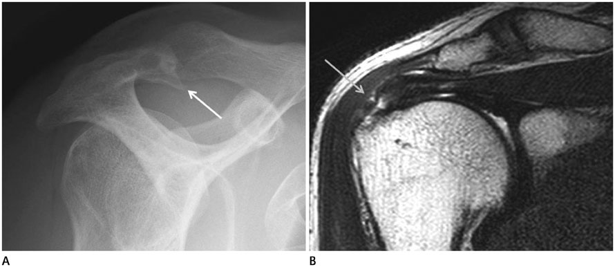
The shoulder radiographs of 104 patients with an arthroscopically proven rotator cuff tear were compared with similar radiographs of 54 age-matched controls with intact cuffs. Two radiologists independently interpreted all radiographs for; cortical thickening with subcortical sclerosis, subcortical cysts, osteophytes in the humeral greater tuberosity, humeral migration, degenerations of the acromioclavicular and glenohumeral joints, and subacromial spurs. Statistical analysis was performed to determine relationships between each type of rotator cuff tears and radiographic abnormalities. Inter-observer agreements with respect to radiographic findings were analyzed.
RESULTS
Humeral migration and degenerative change of the greater tuberosity, including sclerosis, subcortical cysts, and osteophytes, were more associated with full-thickness tears (p < 0.01). Subacromial spurs were more common for full-thickness and bursal-sided tears (p < 0.01). No association was found between degeneration of the acromioclavicular or glenohumeral joint and the presence of a cuff tear.
CONCLUSION
Different types of rotator cuff tears are associated with different radiographic abnormalities.
Figure
Reference
-
1. Hamada K, Fukuda H, Mikasa M, Kobayashi Y. Roentgenographic findings in massive rotator cuff tears. A long-term observation. Clin Orthop Relat Res. 1990; (254):92–96.2. Neer CS 2nd. Impingement lesions. Clin Orthop Relat Res. 1983; (173):70–77.3. Tuite MJ, Toivonen DA, Orwin JF, Wright DH. Acromial angle on radiographs of the shoulder: correlation with the impingement syndrome and rotator cuff tears. AJR Am J Roentgenol. 1995; 165:609–613.4. Norwood LA, Barrack R, Jacobson KE. Clinical presentation of complete tears of the rotator cuff. J Bone Joint Surg Am. 1989; 71:499–505.5. Pearsall AW 4th, Bonsell S, Heitman RJ, Helms CA, Osbahr D, Speer KP. Radiographic findings associated with symptomatic rotator cuff tears. J Shoulder Elbow Surg. 2003; 12:122–127.6. Choi JY, Yum JK, Song MC. Correlation between degree of torn rotator cuff in MRI and degenerative change of acromion and greater tuberosity in simple radiography. Clin Shoulder Elbow. 2010; 16:1–9.7. Saupe N, Pfirrmann CW, Schmid MR, Jost B, Werner CM, Zanetti M. Association between rotator cuff abnormalities and reduced acromiohumeral distance. AJR Am J Roentgenol. 2006; 187:376–382.8. Lee KR, Park JS, Jin WJ, Lee YG, Ryu KN. Plain radiographic findings of a supraspinatus tear: correlation with MR findings. J Korean Radiol Soc. 2007; 57:377–384.9. Huang LF, Rubin DA, Britton CA. Greater tuberosity changes as revealed by radiography: lack of clinical usefulness in patients with rotator cuff disease. AJR Am J Roentgenol. 1999; 172:1381–1388.10. Chopp JN, O'Neill JM, Hurley K, Dickerson CR. Superior humeral head migration occurs after a protocol designed to fatigue the rotator cuff: a radiographic analysis. J Shoulder Elbow Surg. 2010; 19:1137–1144.11. Bonsell S, Pearsall AW 4th, Heitman RJ, Helms CA, Major NM, Speer KP. The relationship of age, gender, and degenerative changes observed on radiographs of the shoulder in asymptomatic individuals. J Bone Joint Surg Br. 2000; 82:1135–1139.
- Full Text Links
- Actions
-
Cited
- CITED
-
- Close
- Share
- Similar articles
-
- Delaminated Rotator Cuff Tear: Concurrent Concept and Treatment
- Biological Characteristics of Rotator Cuff Tendon
- Isolated Medial Dislocation of the Long Head of the Biceps without Rotator Cuff Tear: A Case Report
- The Correlation Between Clinical Features and Radiographic Grades in Massive Rotator Cuff Tear Patients
- Rotator cuff tear with joint stiffness: a review of current treatment and rehabilitation

