J Korean Radiol Soc.
1998 Mar;38(3):531-533. 10.3348/jkrs.1998.38.3.531.
Radiologic Findings of Sinus Pericranii
- Affiliations
-
- 1epartment of Radiology, Neurosurgery, Samsung Medical Center, Sungkyunkwan University College of Medicine.
- 2Department of Radiology Seoul National University College of Medicine.
- KMID: 2201415
- DOI: http://doi.org/10.3348/jkrs.1998.38.3.531
Abstract
- Sinus pericranii is a rare vascular anomaly consisting of abnormal venous communication between intra- andextracranial circulation. We report one case, confirmed by surgery, and describe the radiological findings ofDoppler ultrasonography, CT and MR imaging.
Keyword
Figure
-

Fig. 1. Postcontrast CT with bone window setting shows soft tissue mass (arrowheads) beneath the scalp. Underlying bone erosion with small bone defect (arrow) is also noted.
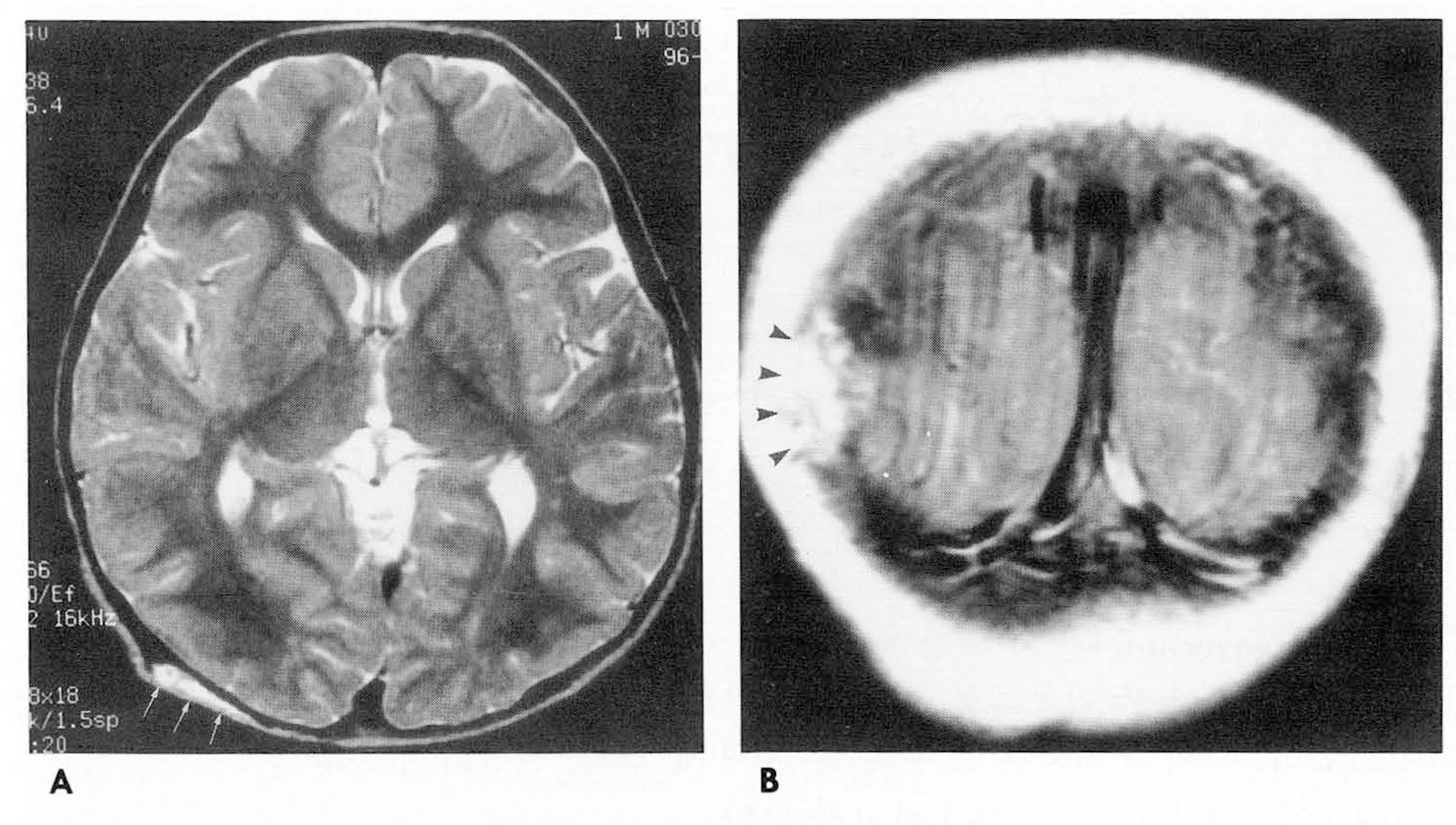
Fig. 2. A. Axial T2-weighted MR image reveals a flat scalp mass of high signal intensity (arrows). There is a suspicious bone erosion in the underlying skull. B. Postcontrast Tl-weighted coronal image demonstrates a well enhancing mass (arrowheads) in right occipital area.
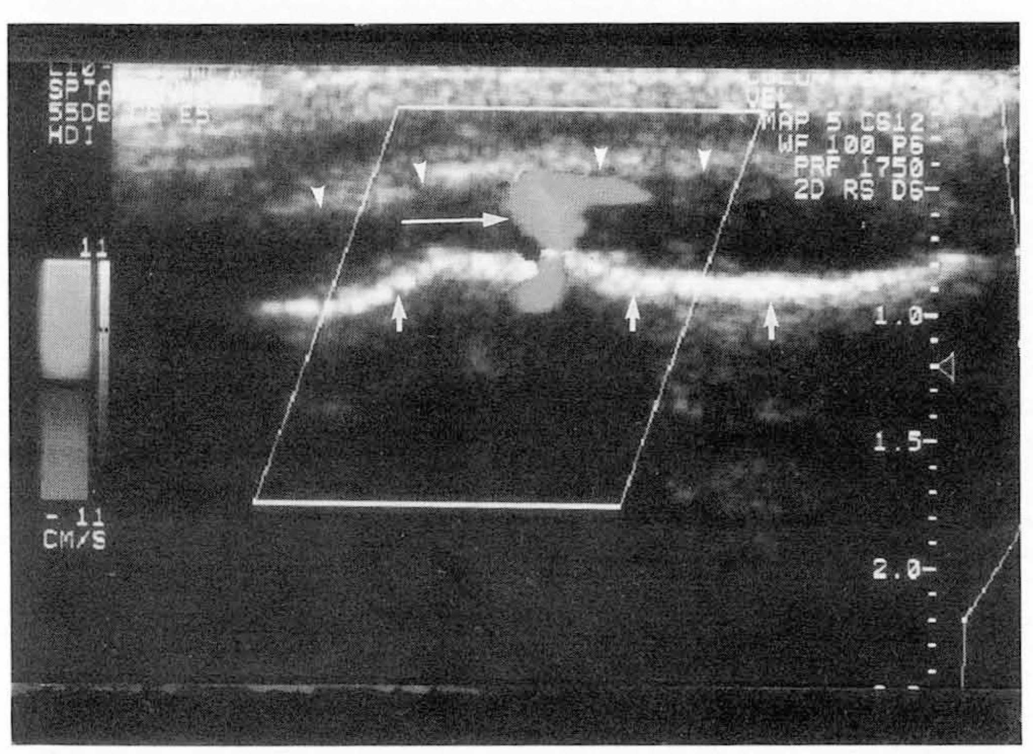
Fig. 3. Color Doppler sonogram shows a diffuse hypoechoic mass (arrowheads) superficial to occipital bone (short arrows). There is an emissary vein (long arrow) in which venous flow was confirmed on pulsed Doppler study (not shown).
Reference
-
1.Sadler LR., Tarr RW., Jungreis CA., Sekhar L. Sinus pericranii: CT and MR findings. J Comput Assist Tomogr. 1990. 14(1):124–127.2.Luker GD., Siegel MJ. Sinus pericranii: sonographic findings. AJR. 1995. 165:175–176.
Article3.Witrak BJ., Davis PC., Hoffman JC Jr. Sinus pericranii. Pediatr Radiol. 1986. 16:55–56.
Article4.Bollar A., Allut AG., Prieto A., Gelabert M., Becerra E. Sinus pericranii: radiological and etiopathological considerations. J Neurosurg. 1992. 77:469–472.
Article
