A case of Paraganglioma Arising in the Transverse Mesocolon
- Affiliations
-
- 1Department of Internal Medicine, Seoul National University College of Medicine, Korea.
- 2Department of Surgery, Seoul National University College of Medicine, Korea.
- KMID: 2200597
- DOI: http://doi.org/10.3803/jkes.2005.20.5.496
Abstract
- Herein, a case of a solitary primary paraganglioma arising in the mesentery, found in a hypertensive 70-year-old woman, who presented with nausea and postprandial abdominal discomfort, is reported. Ultrasonography and computed tomography showed a hypervascular mass abutting the second portion of the duodenum. An exploratory laparotomy revealed a 5.5 x 5.3 x 5cm sized mass in the mesentery of the transverse colon, which was histologically proven to be a paraganglioma. No intraoperative hemodynamic changes developed, and the postoperative course was uneventful. To our knowledge, this is the first case of a paraganglioma arising in the mesentery reported in Korea. Considering the unusual locations and the associated operative risk, it is necessary to rule out the possibility of a functioning paraganglioma in the preoperative differential diagnosis of an abdominal mass.
MeSH Terms
Figure
-
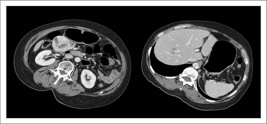
Fig. 1 Computed tomography of abdomen. A 4~5 cm sized, well-enhanced hypervascular mass was detected around the second portion of duodenum. Multiple cystic lesions suspected as metastases were found in the liver segment 8.
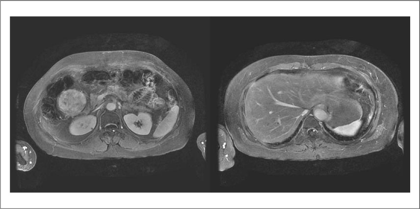
Fig. 2 Magnetic resonance images of the liver. A mass with inhomogenous enhancement abutted on the second portion of duodenum. Multiple cystic lesions in the liver showed low signal intensity after enhancement.
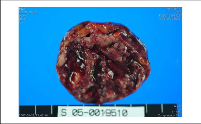
Fig. 3 Gross finding of resected mesenteric mass. A 5.5×5.3×5 cm sized round, slightly hard, well-circumscribed mass was shown here. The findings of hemorrage and necrosis were seen in the cut surface of the mass.
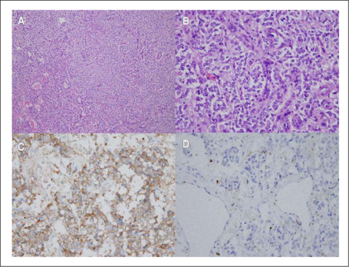
Fig. 4 Microscopic findings of resected mesenteric mass Hematoxylin and eosin stained section showed typical findings of neuroendocrine tumor (A: ×100, B: ×400). Imunohistochemical stain for synaptophysin was positive and that for S-100 was focal positive (C: ×400, D: ×400).
Cited by 1 articles
-
Mesenteric Lesions Appearances on CT with Similar or Distinctive
Hwajin Cha, Jiyoung Hwang, Seong Sook Hong, Eun Ji Lee, Hyun-joo Kim, Yun-Woo Chang
J Korean Soc Radiol. 2019;80(6):1091-1106. doi: 10.3348/jksr.2019.80.6.1091.
Reference
-
1. Lack EE. Extraadrenal paragangliomas of the sympathoadrenal neuroendocrine system. Tumors of the adrenal gland and extra-adrenal paraganglia. 1997. Washington DC: Armed Forces Institute of Pathology;269–282. Atlas of tumor pathology, 3rd series fascicle 19.2. Rosai J. Ackerman's Surgical Pathology. 1997. 8th ed. St Louis: Mol CV Mosby Co;2153–2154.3. Yannopulos K, Stout AP. Primary solid tumors of the mesentery. Cancer. 1963. 16:914–927.4. Glenner GG, Grimley PM. Tumors of the extraadrenal paraganglionic system (including chemoreceptors). Atlas of tumor pathology. 1974. Washington, DC: Armed Forces Institute of Pathology;2nd series, fascicle 9.5. Taylor HB, Helwig EB. Benign non chromaffin paragangliomas of the duodenum. Virchows Arch Pathol Anat. 1962. 335:356–366.6. Elliot GB. Glomus like bodies on the superior mesenteric artery. Can Med Assoc J. 1965. 92:1303–1305.7. Freedman SR, Goldman RL. Normal paraganglion in the mesosigmoid. Hum Pathol. 1981. 12:1037–1038.8. O'Riordain Diarmuid S., Young William F. Jr, Grant Clive S., Carney J. Aidan, van Heerden Jon A. Clinical Spectrum and Outcome of Functional Extraadrenal paraganglioma. World J Surg. 1996. 20:916–922.9. Arean VM, Ramirez de Arellano GA. Intra abdominal nonchromaffin paragangliomas. Ann Surg. 1956. 144:133–137.10. Carmichael JD, Daniel WA, Lamon EW. Mesenteric chemodectoma. Report of a case. Arch Surg. 1970. 101:630–631.11. Tanaka S, Ooshita H, Kaji H. Extraadrenal paraganglioma of the mesenterium (in Japanese). Rinsho Geka. 1991. 46:503–506.12. Onoue S, Katoh T, Chigira H, Matsuo K, Suzuki M, Shibata Y. A case of malignant paraganglioma arising in the mesentery (in Japanese with English abstract). J Jpn Surg Assoc. 1999. 60:3297–3300.13. Jaffer S, Harpaz N. Mesenteric paraganglioma. A case report and review of the literature. Arch Pathol Lab Med. 2002. 126:362–364.14. Muzaffar S, Fatima S, Saddiqui MS, Kayani N, Pervez S, Raja AJ. Mesenteric paraganglioma. Can J Surg. 2002. 45:459–460.15. Canda AE, Sis B, Sokmen S, Fuzun M, Canda MS. An unusual mesent-eric paraganglioma producing human chorionic gona-dotropin. Tumori. 2004. 90:249–252.16. Kudoh A, Tokuhisa Y, Morita K, Hiraki S, Fukuda S, Eguchi N, Iwata T. Mesenteric paraganglioma: Report of a case. Surg Today. 2005. 35:594–597.
- Full Text Links
-
- Actions
-
Cited
- CITED
-
- Close
- Share
-
- Similar articles
-
- Primary Multiple Mesenteric Liposarcoma of the Transverse Mesocolon
- A Case of Pseudocyst Originated from Ectopic Pancreas of Transverse Mesocolon Associated with Colonic Duplication
- A Case of Nonfunctional Paraganglioma of Retroperitonium
- Absence of transverse colon, persistent descending mesocolon, displaced small and large bowels: a rare congenital anomaly with a high risk of volvulus formation
- A Case of Extra-adrenal Paraganglioma of the Scrotum
