J Korean Acad Prosthodont.
2014 Jul;52(3):222-232. 10.4047/jkap.2014.52.3.222.
Comparison of landmark positions between Cone-Beam Computed Tomogram (CBCT) and Adjusted 2D lateral cephalogram
- Affiliations
-
- 1Graduate School of Clinical Dentistry, Ewha Womans University, Seoul, Republic of Korea. minjikim@ewha.ac.kr
- KMID: 2195432
- DOI: http://doi.org/10.4047/jkap.2014.52.3.222
Abstract
- PURPOSE
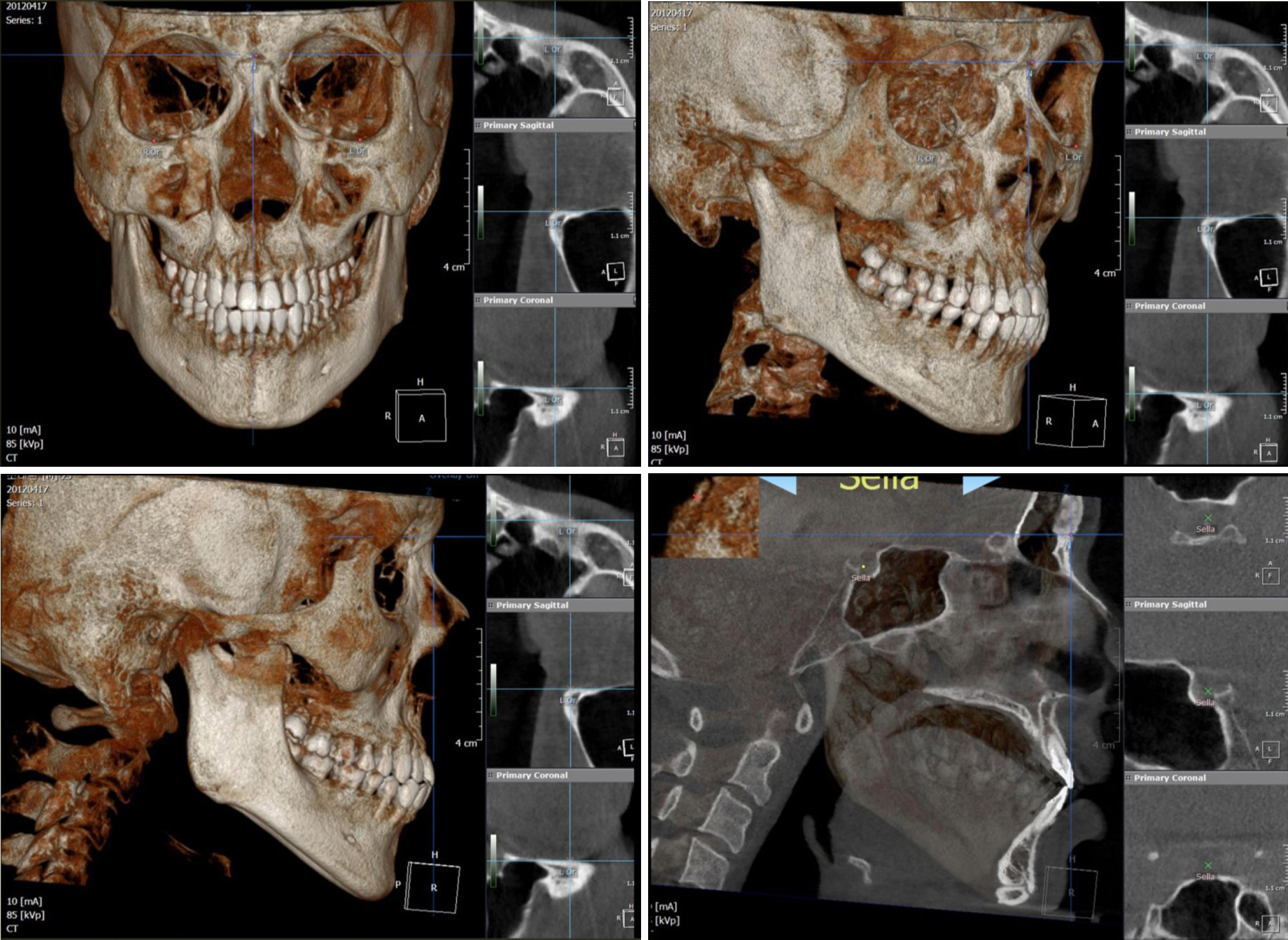
This study aims to investigate if 2D analysis method is applicable to analysis of CBCT by comparing measuring points of CBCT with those of Adjusted 2D Lateral Cephalogram (Adj-Ceph) with magnification adjusted to 100% and finding out at which landmarks the difference in position appear.
MATERIALS AND METHODS
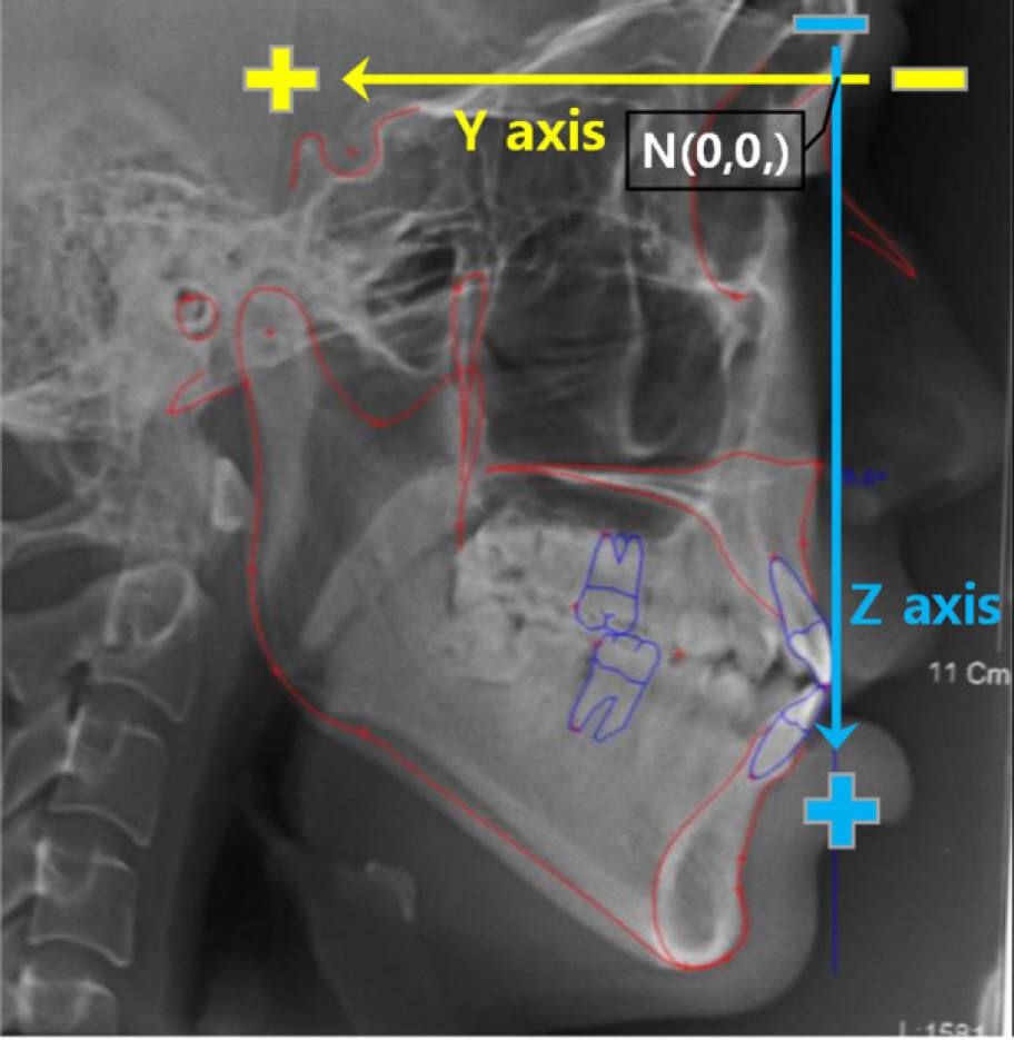
CBCT data and Adj-Ceph (100% magnification) data from 50 adult patients have been extracted as research objects, and the horizontal (Y axis) and vertical (Z axis) coordinates of landmarks were compared. Landmarks have been categorized into 4 groups by the position and whether they are bilaterally overlapped. Paired t-test was used to compare differences between Adj-Ceph and CBCT.
RESULTS
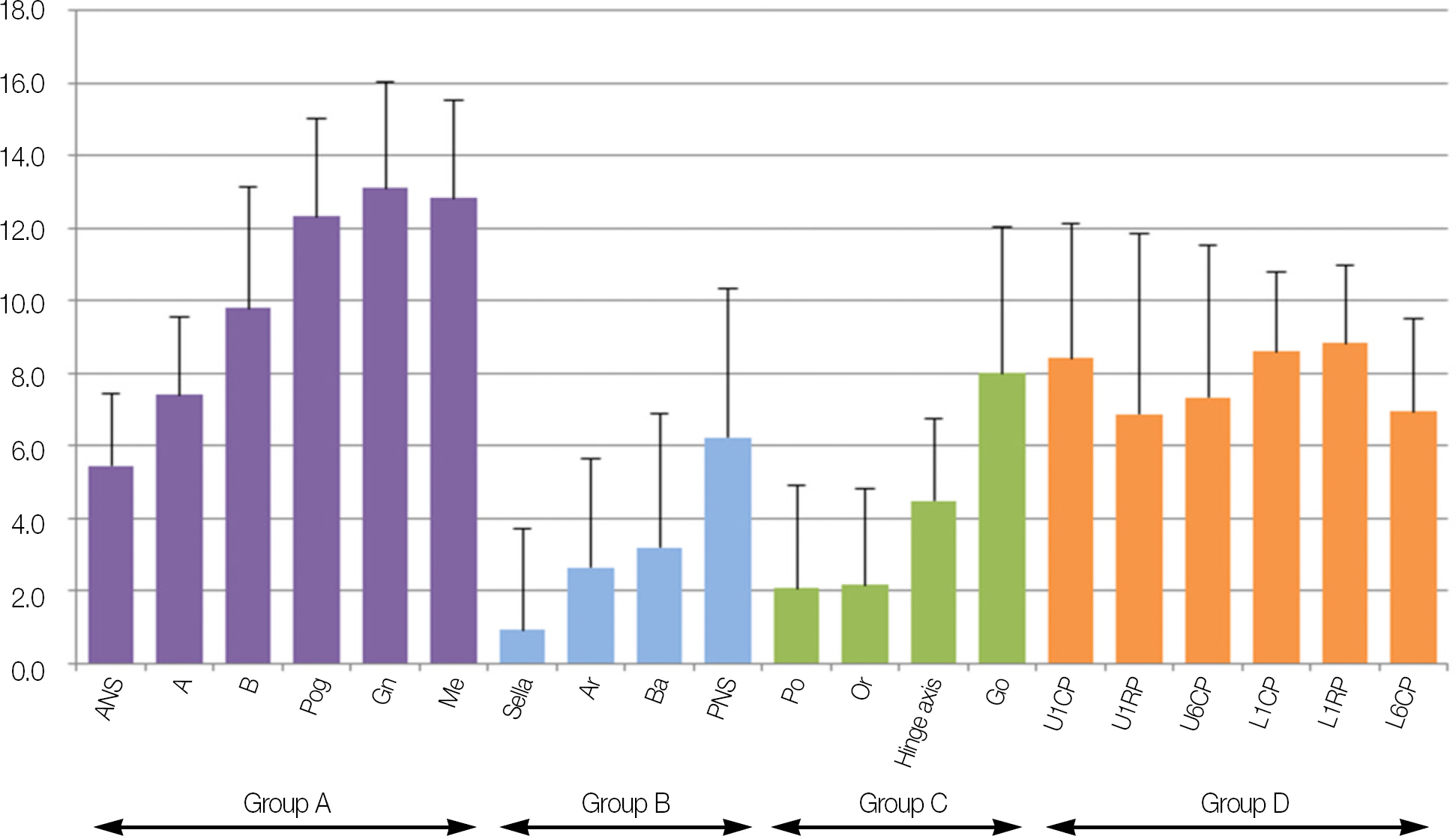
Significant difference was found at 11 landmarks including Group B (S, Ar, Ba, PNS), Group C (Po, Or, Hinge axis, Go) and Group D (U1RP, U6CP, L6CP) in the horizontal (Y) axis while all the landmarks in vertical (Z) axis showed significant difference (P<.05). As a result of landmark difference analysis, a meaningful difference with more than 1 mm at 13 landmarks were indentifed in the horizontal axis. In the vertical axis, significant difference over 1 mm was detected from every landmark except Sella.
CONCLUSION
Using the conventional lateral cephalometric measurements on CBCT is insufficient. A new 3D analysis or a modified 2D analysis adjusted on 19 landmarks of the vertical axis and 13 of the horizontal axis are needed when implementing CBCT diagnosis.
MeSH Terms
Figure
Reference
-
1. Broadbent BH. A new x-ray technique and its application to or-thodontia. Angle Orthod. 1981; 51:93–114.2. Broadbent BH. The face of the normal child. Angle Orthod. 1937; 7:183–208.3. Brodie AG. On the growth pattern of the human head. From the third month to the eighth year of life. Am J Anat. 1941; 68:209–62.
Article4. Salzmann JA. The face in profile: an anthropological x-ray investigation on Swedish children and conscripts by Arne Bjo¨rk. Am J Orthod. 1948; 34:691–9.5. Downs WB. Variations in facial relationships; their significance in treatment and prognosis. Am J Orthod. 1948; 34:812–40.
Article6. Steiner CC. Cephalometrics for you and me. Am J Orthod. 1953; 39:729–55.
Article7. Sassouni V. A roentgenographic cephalometric analysis of cephalo-facio-dental relationships. Am J Orthod. 1955; 41:735–64.
Article8. Tweed CH. Was the development of the diagnostic facial triangle as an accurate analysis based on fact or fancy? Am J Orthod. 1962; 48:823–40.
Article9. Harvold EP. The role of function in the etiology and treatment of malocclusion. Am J Orthod. 1968; 54:883–98.
Article10. Jacobson A. Application of the "Wits" appraisal. Am J Orthod. 1976; 70:179–89.
Article11. Jacobson A. The "Wits" appraisal of jaw disharmony. Am J Orthod. 1975; 67:125–38.
Article12. Burstone CJ, James RB, Legan H, Murphy GA, Norton LA. Cephalometrics for orthognathic surgery. J Oral Surg. 1978; 36:269–77.13. Ricketts RM. Perspectives in the clinical application of cephalometrics. The first fifty years. Angle Orthod. 1981; 51:115–50.14. McNamara JA Jr. A method of cephalometric evaluation. Am J Orthod. 1984; 86:449–69.
Article15. Yen PKJ. Identification Of Landmarks In Cephalometric Radiographs. Angle Orthod. 1960; 30:35–41.16. Marshall D. Interpretation of the posteroanterior skull radi-ograph-assembly of disarticulated bones. Dent Radiogr Photogr. 1969; 42:27–35.17. Baumrind S, Frantz RC. The reliability of head film measurements.1. Landmark identification. Am J Orthod. 1971; 60:111–27.18. Midtga�rd J, Bjo¨rk G, Linder-Aronson S. Reproducibility of cephalometric landmarks and errors of measurements of cephalometric cranial distances. Angle Orthod. 1974; 44:56–61.19. Cho HJ. A three-dimensional cephalometric analysis. J Clin Orthod. 2009; 43:235–52.20. Grayson BH, McCarthy JG, Bookstein F. Analysis of craniofacial asymmetry by multiplane cephalometry. Am J Orthod. 1983; 84:217–24.
Article21. Baumrind S, Moffitt FH, Curry S. Three-dimensional x-ray stereometry from paired coplanar images: a progress report. Am J Orthod. 1983; 84:292–312.
Article22. Kusnoto B, Evans CA, BeGole EA, de Rijk W. Assessment of 3-dimensional computer-generated cephalometric measurements. Am J Orthod Dentofacial Orthop. 1999; 116:390–9.
Article23. Dale AM, Robert AD. A Clinician's Guide to Understanding Cone Beam Volumetric Imaging (CBVI). 2007. [cited 2012 December 20]. Available from:. http://www.Ineedce.com/courses/1413/PDF/A_Clin_Gde_ConeBeam.pdf.24. Cavalcanti MG, Vannier MW. Quantitative analysis of spiral computed tomography for craniofacial clinical applications. Dentomaxillofac Radiol. 1998; 27:344–50.
Article25. Matteson SR, Bechtold W, Phillips C, Staab EV. A method for three-dimensional image reformation for quantitative cephalometric analysis. J Oral Maxillofac Surg. 1989; 47:1053–61.
Article26. Christiansen EL, Thompson JR, Kopp S. Intra- and inter-observer variability and accuracy in the determination of linear and angular measurements in computed tomography. An in vitro and in situ study of human mandibles. Acta Odontol Scand. 1986; 44:221–9.
Article27. Hildebolt CF, Vannier MW, Knapp RH. Validation study of skull three-dimensional computerized tomography measurements. Am J Phys Anthropol. 1990; 82:283–94.
Article28. Lascala CA, Panella J, Marques MM. Analysis of the accuracy of linear measurements obtained by cone beam computed tomography (CBCT-NewTom). Dentomaxillofac Radiol. 2004; 33:291–4.
Article29. Schlicher W, Nielsen I, Huang JC, Maki K, Hatcher DC, Miller AJ. Consistency and precision of landmark identification in three-dimensional cone beam computed tomography scans. Eur J Orthod. 2012; 34:263–75.
Article30. Grauer D, Cevidanes LS, Styner MA, Heulfe I, Harmon ET, Zhu H, Proffit WR. Accuracy and landmark error calculation using cone-beam computed tomography-generated cephalograms. Angle Orthod. 2010; 80:286–94.
Article31. Park JW, Kim NK, Chang YI. Comparison of landmark position between conventional cephalometric radiography and CT scans projected to midsagittal plane. Korean J Orthod. 2008; 38:427–36.
Article32. Kumar V, Ludlow JB, Mol A, Cevidanes L. Comparison of conventional and cone beam CT synthesized cephalograms. Dentomaxillofac Radiol. 2007; 36:263–9.
Article33. Kumar V, Ludlow J, Soares Cevidanes LH, Mol A. In vivo comparison of conventional and cone beam CT synthesized cephalograms. Angle Orthod. 2008; 78:873–9.
Article34. Terajima M, Yanagita N, Ozeki K, Hoshino Y, Mori N, Goto TK, Tokumori K, Aoki Y, Nakasima A. Three-dimensional analysis system for orthognathic surgery patients with jaw deformities. Am J Orthod Dentofacial Orthop. 2008; 134:100–11.
Article35. Terajima M, Endo M, Aoki Y, Yuuda K, Hayasaki H, Goto TK, Tokumori K, Nakasima A. Four-dimensional analysis of stom-atognathic function. Am J Orthod Dentofacial Orthop. 2008; 134:276–87.
Article36. Suri S, Utreja A, Khandelwal N, Mago SK. Craniofacial computerized tomography analysis of the midface of patients with re-paired complete unilateral cleft lip and palate. Am J Orthod Dentofacial Orthop. 2008; 134:418–29.
Article37. Kau CH, Richmond S. Three-dimensional analysis of facial morphology surface changes in untreated children from 12 to 14 years of age. Am J Orthod Dentofacial Orthop. 2008; 134:751–60.
Article38. Garrett BJ, Caruso JM, Rungcharassaeng K, Farrage JR, Kim JS, Taylor GD. Skeletal effects to the maxilla after rapid maxillary expansion assessed with cone-beam computed tomography. Am J Orthod Dentofacial Orthop. 2008; 134:8–9.
Article39. Phatouros A, Goonewardene MS. Morphologic changes of the palate after rapid maxillary expansion: a 3-dimensional computed tomography evaluation. Am J Orthod Dentofacial Orthop. 2008; 134:117–24.
Article40. Ballanti F, Lione R, Fanucci E, Franchi L, Baccetti T, Cozza P. Immediate and post-retention effects of rapid maxillary expansion investigated by computed tomography in growing patients. Angle Orthod. 2009; 79:24–9.
Article41. Kragskov J, Bosch C, Gyldensted C, Sindet-Pedersen S. Comparison of the reliability of craniofacial anatomic landmarks based on cephalometric radiographs and three-dimensional CT scans. Cleft Palate Craniofac J. 1997; 34:111–6.
Article42. Landis JR, Koch GG. The measurement of observer agree-ment for categorical data. Biometrics. 1977; 33:159–74.
Article43. Kim JY, Lee DK, Lee SH. Comparison of the observer reliability of cranial anatomic landmarks based on cephalometric radiograph and three-dimensional computed tomography scans. J Korean Assoc Oral Maxillofac Surg. 2010; 36:262–9.
Article44. van Vlijmen OJ, Maal TJ, Berge′ SJ, Bronkhorst EM, Katsaros C, Kuijpers-Jagtman AM. A comparison between two-dimensional and three-dimensional cephalometry on frontal radiographs and on cone beam computed tomography scans of human skulls. Eur J Oral Sci. 2009; 117:300–5.
Article45. Adams GL, Gansky SA, Miller AJ, Harrell WE Jr, Hatcher DC. Comparison between traditional 2-dimensional cephalometry and a 3-dimensional approach on human dry skulls. Am J Orthod Dentofacial Orthop. 2004; 126:397–409.
Article
- Full Text Links
- Actions
-
Cited
- CITED
-
- Close
- Share
- Similar articles
-
- The reliability of the cephalogram generated from cone-beam CT
- A comparative study of the reproducibility of landmark identification on posteroanterior and anteroposterior cephalograms generated from cone-beam computed tomography scans
- Accuracy and reproducibility of landmark of cone beam computed tomography (CT) synthesized cephalograms
- Use of Reference Ear Plug to improve accuracy of lateral cephalograms generated from cone-beam computed tomography scans
- Comparison of cone-beam computed tomography cephalometric measurements using a midsagittal projection and conventional two-dimensional cephalometric measurements


