J Korean Med Assoc.
2013 Nov;56(11):1017-1022. 10.5124/jkma.2013.56.11.1017.
Prevention and treatment of hallux valgus
- Affiliations
-
- 1Department of Orthopaedic Surgery, Seoul National University Boramae Medical Center, Seoul, Korea. kjh12344@hanmail.net
- KMID: 2193477
- DOI: http://doi.org/10.5124/jkma.2013.56.11.1017
Abstract
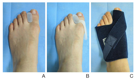
- In hallux valgus, one of the most common conditions affecting the forefoot, the first metatarsophalangeal joint is progressively subluxed due to lateral deviation of the hallux and medial deviation of the first metatarsal. Patients usually complain of medial prominence pain, commonly referred to as "bunion pain," plantar keratotic lesions, and lesser toe deformities such as hammer toe or claw toe deformities. The etiology of hallux valgus is multifactorial. Narrow high-heeled shoes or excessive weight-bearing have been suggested to be extrinsic factors contributing to the condition, and many other intrinsic factors also exist, such as genetics, ligamentous laxity, metatarsus primus varus, pes planus, functional hallux limitus, sexual dimorphism, age, metatarsal morphology, first-ray hypermobility, and tight Achilles tendon. When we evaluate patients with hallux valgus, careful history taking and meticulous examination are necessary. On the radiographic evaluation, we routinely measure the hallux valgus angle, intermetatarsal angle, and distal metatarsal articular angle, which are valuable parameters in decision making for bunion surgery. To prevent the development and progression of hallux valgus, a soft leather shoe with a wide toe box is usually recommended. The use of a toe separator or bunion splint may help in relieving symptoms. The purpose of hallux valgus surgery is to correct the deformity and maintain a biomechanically functional foot. When we decide on an adequate surgical option, we should consider the patient's subjective symptoms, the expectations of the patient, the degree of the de-formity, and the radiographic measurements in order to correct the deformity and prevent complications after surgery.
Keyword
MeSH Terms
Figure
Cited by 1 articles
-
Relationship between Bony Alignment of Foot and Scoliosis in Children and Adolescent
Jae Hwang Song, Woo Jin Shin, Sung Jun Moon, Jin Woong Yi, Tae Gyun Kim
J Korean Foot Ankle Soc. 2024;28(2):48-54. doi: 10.14193/jkfas.2024.28.2.48.
Reference
-
1. Vanore JV, Christensen JC, Kravitz SR, Schuberth JM, Thomas JL, Weil LS, Zlotoff HJ, Mendicino RW, Couture SD. Clinical Practice Guideline First Metatarsophalangeal Joint Disorders Panel of the American College of Foot and Ankle Surgeons. Diagnosis and treatment of first metatarsophalangeal joint disorders. Section 1: hallux valgus. J Foot Ankle Surg. 2003; 42:112–123.
Article2. Menz HB, Lord SR. Gait instability in older people with hallux valgus. Foot Ankle Int. 2005; 26:483–489.
Article3. Tinetti ME, Speechley M, Ginter SF. Risk factors for falls among elderly persons living in the community. N Engl J Med. 1988; 319:1701–1707.
Article4. Nix S, Smith M, Vicenzino B. Prevalence of hallux valgus in the general population: a systematic review and meta-analysis. J Foot Ankle Res. 2010; 3:21.
Article5. Roddy E, Zhang W, Doherty M. Prevalence and associations of hallux valgus in a primary care population. Arthritis Rheum. 2008; 59:857–862.
Article6. Nguyen US, Hillstrom HJ, Li W, Dufour AB, Kiel DP, Procter-Gray E, Gagnon MM, Hannan MT. Factors associated with hallux valgus in a population-based study of older women and men: the MOBILIZE Boston Study. Osteoarthritis Cartilage. 2010; 18:41–46.
Article7. Kato T, Watanabe S. The etiology of hallux valgus in Japan. Clin Orthop Relat Res. 1981; (157):78–81.
Article8. Corrigan JP, Moore DP, Stephens MM. Effect of heel height on forefoot loading. Foot Ankle. 1993; 14:148–152.
Article9. Cho NH, Kim S, Kwon DJ, Kim HA. The prevalence of hallux valgus and its association with foot pain and function in a rural Korean community. J Bone Joint Surg Br. 2009; 91:494–498.
Article10. Perera AM, Mason L, Stephens MM. The pathogenesis of hallux valgus. J Bone Joint Surg Am. 2011; 93:1650–1661.
Article11. Maclennan R. Prevalence of hallux valgus in a neolithic New Guinea population. Lancet. 1966; 1:1398–1400.
Article12. Hewitt D, Stewart AM, Webb JW. The prevalence of foot defects among wartime recruits. Br Med J. 1953; 2:745–749.
Article13. Coughlin MJ, Jones CP. Hallux valgus: demographics, etiology, and radiographic assessment. Foot Ankle Int. 2007; 28:759–777.
Article14. Pique-Vidal C, Sole MT, Antich J. Hallux valgus inheritance: pedigree research in 350 patients with bunion deformity. J Foot Ankle Surg. 2007; 46:149–154.
Article15. Saro C, Andren B, Wildemyr Z, Fellander-Tsai L. Outcome after distal metatarsal osteotomy for hallux valgus: a prospective randomized controlled trial of two methods. Foot Ankle Int. 2007; 28:778–787.
Article16. Frey C, Thompson F, Smith J, Sanders M, Horstman H. American Orthopaedic Foot and Ankle Society women's shoe survey. Foot Ankle. 1993; 14:78–81.
Article17. Carl A, Ross S, Evanski P, Waugh T. Hypermobility in hallux valgus. Foot Ankle. 1988; 8:264–270.
Article18. Scott G, Menz HB, Newcombe L. Age-related differences in foot structure and function. Gait Posture. 2007; 26:68–75.
Article19. Humbert JL, Bourbonniere C, Laurin CA. Metatarsophalangeal fusion for hallux valgus: indications and effect on the first metatarsal ray. Can Med Assoc J. 1979; 120:937–941. 95620. Heden RI, Sorto LA Jr. The Buckle point and the metatarsal protrusion's relationship to hallux valgus. J Am Podiatry Assoc. 1981; 71:200–208.
Article21. Wilson DW. Treatment of hallux valgus and bunions. Br J Hosp Med. 1980; 24:548–549.22. Coughlin MJ, Thompson FM. The high price of high-fashion footwear. Instr Course Lect. 1995; 44:371–377.23. Tehraninasr A, Saeedi H, Forogh B, Bahramizadeh M, Keyhani MR. Effects of insole with toe-separator and night splint on patients with painful hallux valgus: a comparative study. Prosthet Orthot Int. 2008; 32:79–83.
Article24. Budiman-Mak E, Conrad KJ, Roach KE, Moore JW, Lertratanakul Y, Koch AE, Skosey JL, Froelich C, Joyce-Clark N. Can foot orthoses prevent hallux valgus deformity in rheumatoid arthritis?: a randomized clinical trial. J Clin Rheumatol. 1995; 1:313–322.
Article25. Geissele AE, Stanton RP. Surgical treatment of adolescent hallux valgus. J Pediatr Orthop. 1990; 10:642–648.
Article26. Grill F, Hetherington V, Steinbock G, Altenhuber J. Experiences with the chevron (V-)osteotomy on adolescent hallux valgus. Arch Orthop Trauma Surg. 1986; 106:47–51.27. Mann RA. Decision-making in bunion surgery. Instr Course Lect. 1990; 39:3–13.28. Lee WC, Kim YM. Correction of hallux valgus using lateral soft-tissue release and proximal Chevron osteotomy through a medial incision. J Bone Joint Surg Am. 2007; 89:Suppl 3. 82–89.
Article29. Park JY, Jung HG, Kim TH, Kang MS. Intraoperative incidence of hallux valgus interphalangeus following basilar first metatarsal osteotomy and distal soft tissue realignment. Foot Ankle Int. 2011; 32:1058–1062.
Article30. Richardson EG. Complications after hallux valgus surgery. Instr Course Lect. 1999; 48:331–342.
- Full Text Links
- Actions
-
Cited
- CITED
-
- Close
- Share
- Similar articles
-
- Incidence of Hallux Valgus Interphalangeus in the Normal and Hallux Valgus Feet and its Correlations with Hallux Valgus Angle and Intermetatarsal Angle
- Consideration of Various Medial Capsulorrhaphy Methods in Hallux Valgus Surgery
- Corrective Osteotomies in Hallux Valgus
- Technique Tip: A Simple Method to Treat Hallux Valgus with Severe Metatarsus Adductus
- A clinical analysis of the operative treatment in hallux valgus


