J Korean Neurosurg Soc.
2016 Mar;59(2):168-171. 10.3340/jkns.2016.59.2.168.
Growing Hemorrhagic Choroidal Fissure Cyst
- Affiliations
-
- 1Department of Neurosurgery, Izmir Katip Celebi University Ataturk Education and Research Hospital, Izmir, Turkey. aysekaratas@yahoo.com
- 2Department of Radiology, Izmir Katip Celebi University Ataturk Education and Research Hospital, Izmir, Turkey.
- KMID: 2192050
- DOI: http://doi.org/10.3340/jkns.2016.59.2.168
Abstract
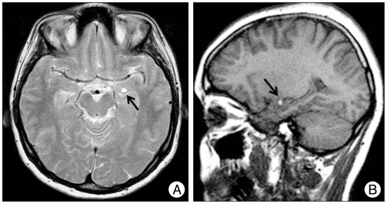
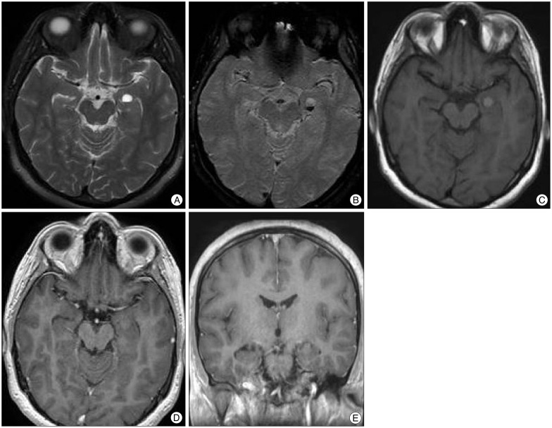
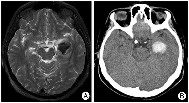
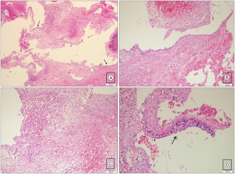
- Choroidal fissure cysts are often incidentally discovered. They are usually asymptomatic. The authors report a case of growing and hemorrhagic choroidal fissure cyst which was treated surgically. A 22-year-old female presented with headache. Cranial MRI showed a left-sided choroidal fissure cyst. Follow-up MRI showed that the size of the cyst had increased gradually. Twenty months later, the patient was admitted to our emergency department with severe headache. MRI and CT showed an intracystic hematoma. Although such cysts usually have a benign course without symptoms and progression, they may rarely present with intracystic hemorrhage, enlargement of the cyst and increasing symptomatology.
Keyword
MeSH Terms
Figure
Reference
-
1. Baka JJ, Sanders WP. MRI of hemorrhagic choroid plexus cyst. Neuroradiology. 1993; 35:428–430. PMID: 8377913.
Article2. de Jong L, Thewissen L, van Loon J, Van Calenbergh F. Choroidal fissure cerebrospinal fluid-containing cysts : case series, anatomical consideration, and review of the literature. World Neurosurg. 2011; 75:704–708. PMID: 21704940.
Article3. Guermazi A, Miaux Y, Majoulet JF, Lafitte F, Chiras J. Imaging findings of central nervous system neuroepithelial cysts. Eur Radiol. 1998; 8:618–623. PMID: 9569335.
Article4. Rhoton AL Jr. The lateral and third ventricles. Neurosurgery. 2002; 51(4 Suppl):S207–S271. PMID: 12234450.
Article


