Cauda Equina Syndrome Associated with Dural Ectasia in Chronic Anlylosing Spondylitis
- Affiliations
-
- 1Department of Neurosurgery, Seoul St. Mary's Hospital, College of Medicine, The Catholic University of Korea, Seoul, Korea. sbc@catholic.ac.kr
- 2The Catholic Neuroscience Institute, College of Medicine, The Catholic University of Korea, Seoul, Korea.
- KMID: 2191150
- DOI: http://doi.org/10.3340/jkns.2014.56.6.517
Abstract
- Cauda equina syndrome (CES) associated with dural ectasia is a rare neurologic complication in patients with longstanding ankylosing spondylitis (AS). We report a 68-year-old male with a 30-year history of AS who presented a typical symptom and signs of progressive CES, urinary incontinence and neuropathic pain of the lumbosacral radiculopathy. Computed tomography (CT) and magnetic resonance imaging (MRI) findings showed the unique appearances of dural ectasia, multiple dural diverticula, erosion of posterior element of the lumbar spine, tethering of the conus medullaris and adhesion of the lumbosacral nerve roots to the posterior aspect of the dural ectasia. Considering the progressive worsening of the clinical signs, detethering of the conus medullaris through resection of the filum terminale was performed through a limited laminectomy. However, the urinary incontinence did not improve and there was a partial relief of the neuropathic leg pain only. The possible pathogenetic mechanism of CES-AS and the dural ectasia in this patient with longstanding AS are discussed with a literature review.
MeSH Terms
Figure
-
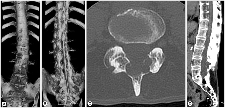
Fig. 1 A : A three-dimensional (3D) reconstructed computed tomography (CT) showing a typical appearance of 'bamboo spine' of ankylosing spondylitis in the anterior view. B : A posterior view on a 3D CT scan showing the fusion of sacroiliac joints, syndesmophyte formation and also multiple erosions of the roof of an enlarged spinal canal. An axial (C) and sagittal CT (D) scan images showing a widened spinal canal, multiple erosions on laminae and spinous process, and a subarachnoid diverticulum protruded through the right intervertebral foramen.

Fig. 2 A : A sagittal T2-weighted magnetic resonance (MR) image showing the low-lying conus medullaris at the level of L2 and an adhesion of the lumbosacral nerve root to the dural ectasia. B : An axial T2-weighted MR image showing multiple dural diverticulas and an adhesion of the nerve root to the posterior wall of the dural diverticula.
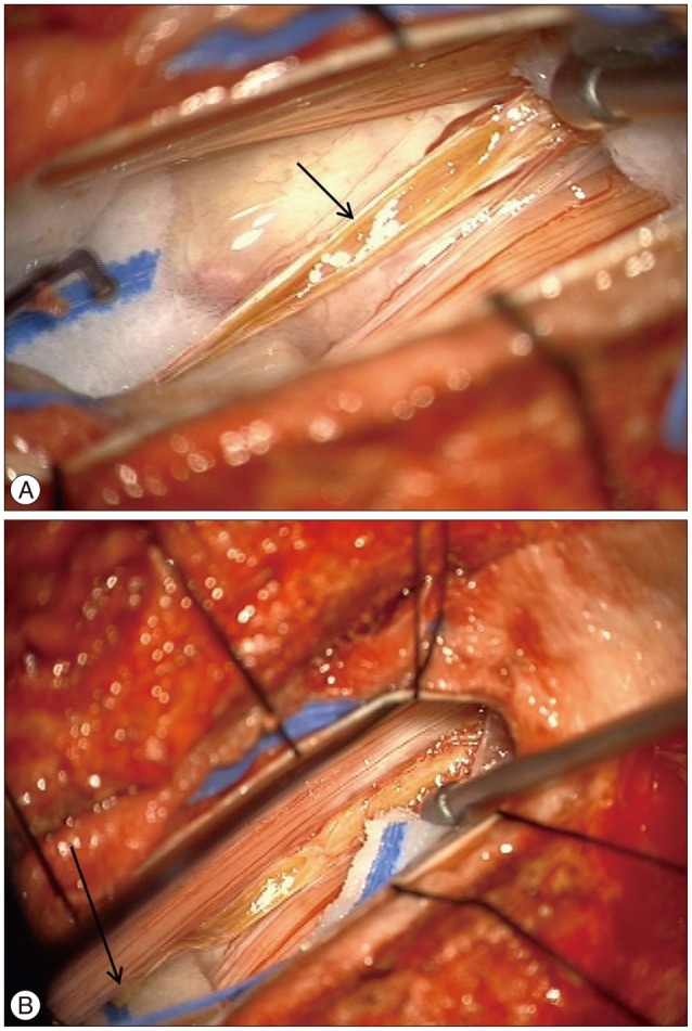
Fig. 3 A : An intraoperative photograph showing the filum terminale through a L2 laminectomy. Note the characteristic fatty infiltration in the filum terminale (arrow). B : An interoperative photography showing the dethetering of the filum terminale after coagulation and cutting (the arrow indicates the cut edge of the filum).
Reference
-
1. Abelló R, Rovira M, Sanz MP, Gili J, Capdevila A, Escalada J, et al. MRI and CT of ankylosing spondylitis with vertebral scalloping. Neuroradiology. 1988; 30:272–275. PMID: 3405419.
Article2. Ahn NU, Ahn UM, Nallamshetty L, Springer BD, Buchowski JM, Funches L, et al. Cauda equina syndrome in ankylosing spondylitis (the CES-AS syndrome) : meta-analysis of outcomes after medical and surgical treatments. J Spinal Disord. 2001; 14:427–433. PMID: 11586143.
Article3. Arslanoglu A, Aygun N. Magnetic resonance imaging of cauda equina syndrome in long-standing ankylosing spondylitis. Australas Radiol. 2007; 51:375–377. PMID: 17635477.
Article4. Bartleson JD, Cohen MD, Harrington TM, Goldstein NP, Ginsburg WW. Cauda equina syndrome secondary to long-standing ankylosing spondylitis. Ann Neurol. 1983; 14:662–669. PMID: 6651250.
Article5. Bowie EA, Glasgow GL. Cauda equina lesions associated with ankylosing spondylitis. Br Med J. 1961; 2:24–27. PMID: 20789178.
Article6. Confavreux C, Larbre JP, Lejeune E, Sindou M, Aimard G. Cerebrospinal fluid dynamics in the tardive cauda equina syndrome of ankylosing spondylitis. Ann Neurol. 1991; 29:221–223. PMID: 2012391.
Article7. Cornec D, Devauchelle Pensec V, Joulin SJ, Saraux A. Dramatic efficacy of infliximab in cauda equina syndrome complicating ankylosing spondylitis. Arthritis Rheum. 2009; 60:1657–1660. PMID: 19479855.
Article8. Dinichert A, Cornelius JF, Lot G. Lumboperitoneal shunt for treatment of dural ectasia in ankylosing spondylitis. J Clin Neurosci. 2008; 15:1179–1182. PMID: 18710808.
Article9. Hauge T. Chronic rheumatoid polyarthritis and spondyl-arthritis associated with neurological symptoms and signs occasionally simulating an intraspinal expansive process. Acta Chir Scand. 1961; 120:395–401. PMID: 13712291.10. Lan HH, Chen DY, Chen CC, Lan JL, Hsieh CW. Combination of transverse myelitis and arachnoiditis in cauda equina syndrome of long-standing ankylosing spondylitis : MRI features and its role in clinical management. Clin Rheumatol. 2007; 26:1963–1967. PMID: 17332972.
Article11. Larner AJ, Pall HS, Hockley AD. Arrested progression of the cauda equina syndrome of ankylosing spondylitis after lumboperitoneal shunting. J Neurol Neurosurg Psychiatry. 1996; 61:115–116. PMID: 8676141.
Article12. Liu CC, Lin YC, Lo CP, Chang TP. Cauda equina syndrome and dural ectasia : rare manifestations in chronic ankylosing spondylitis. Br J Radiol. 2011; 84:e123–e125. PMID: 21606066.13. Matthews WB. The neurological complications of ankylosing spondylitis. J Neurol Sci. 1968; 6:561–573. PMID: 4303835.
Article14. Nallamshetty L, Ahn NU, Ahn UM, Nallamshetty HS, Rose PS, Buchowski JM, et al. Dural ectasia and back pain : review of the literature and case report. J Spinal Disord Tech. 2002; 15:326–329. PMID: 12177551.15. Rubenstein DJ, Alvarez O, Ghelman B, Marchisello P. Cauda equina syndrome complicating ankylosing spondylitis : MR features. J Comput Assist Tomogr. 1989; 13:511–513. PMID: 2723187.16. Shaw PJ, Allcutt DA, Bates D, Crawford PJ. Cauda equina syndrome associated with multiple lumbar arachnoid cysts in ankylosing spondylitis : improvement following surgical therapy. J Neurol Neurosurg Psychiatry. 1990; 53:1076–1079. PMID: 2292702.
Article17. Soeur M, Monseu G, Baleriaux-Waha D, Duchateau M, Williame E, Pa-steels JL. Cauda equina syndrome in ankylosing spondylitis. Anatomical, diagnostic, and therapeutic considerations. Acta Neurochir (Wien). 1981; 55:303–315. PMID: 7234535.18. Sparling MJ, Bartleson JD, McLeod RA, Cohen MD, Ginsburg WW. Magnetic resonance imaging of arachnoid diverticula associated with cauda equina syndrome in ankylosing spondylitis. J Rheumatol. 1989; 16:1335–1337. PMID: 2509695.19. Tallroth K, Malmivaara A, Laitinen ML, Savolainen A, Harilainen A. Lum-bar spine in Marfan syndrome. Skeletal Radiol. 1995; 24:337–340. PMID: 7570153.
Article
- Full Text Links
-
- Actions
-
Cited
- CITED
-
- Close
- Share
-
- Similar articles
-
- MRI of Cauda Equina Syndrome in Ankylosing Spondylitis: A Case Report
- Marfan syndrome and symptomatic dural ectasia: A case report and literature review
- Causes and Clinical Manifestations of Cauda Equina Syndrome
- Cavernous Hemangioma of the Cauda Equina
- Treatment of a 3rd Lumbar Vertebra Translational Injury Combined with Incomplete Cauda Equina Syndrome in Ankylosing Spondylitis: A Case Report
