Reliability of Stereotactic Coordinates of 1.5-Tesla and 3-Tesla MRI in Radiosurgery and Functional Neurosurgery
- Affiliations
-
- 1Gamma Knife Center, Haeundae Paik Hospital, Inje University College of Medicine, Busan, Korea. hykim0803@naver.com
- 2Department of Neurosurgery, Haeundae Paik Hospital, Inje University College of Medicine, Busan, Korea.
- KMID: 2191078
- DOI: http://doi.org/10.3340/jkns.2014.55.3.136
Abstract
OBJECTIVE
The aims of this study are to identify interpersonal differences in defining coordinates and to figure out the degree of distortion of the MRI and compare the accuracy between CT, 1.5-tesla (T) and 3.0T MRI.
METHODS
We compared coordinates in the CT images defined by 2 neurosurgeons. We also calculated the errors of 1.5T MRI and those of 3.0T. We compared the errors of the 1.5T with those of the 3.0T. In addition, we compared the errors in each sequence and in each axis.
RESULTS
The mean difference in the CT images between the two neurosurgeons was 0.48+/-0.22 mm. The mean errors of the 1.5T were 1.55+/-0.48 mm (T1), 0.75+/-0.38 (T2), and 1.07+/-0.57 (FLAIR) and those of the 3.0T were 2.35+/-0.53 (T1), 2.18+/-0.76 (T2), and 2.16+/-0.77 (FLAIR). The smallest mean errors out of all the axes were in the x axis : 0.28-0.34 (1.5T) and 0.31-0.52 (3.0T). The smallest errors out of all the MRI sequences were in the T2 : 0.29-0.58 (1.5T) and 0.31-1.85 (3.0T).
CONCLUSION
There was no interpersonal difference in running the Gamma Plan(R) to define coordinates. The errors of the 3.0T were greater than those of the 1.5T, and these errors were not of an acceptable level. The x coordinate error was the smallest and the z coordinate error was the greatest regardless of the MRI sequence. The T2 sequence was the most accurate sequence.
Figure
-
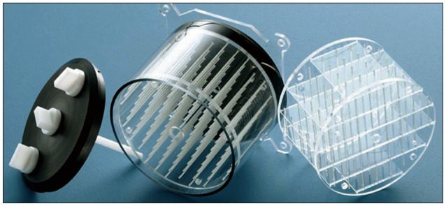
Fig. 1 The Elekta magnetic resonance imaging phantom was used in this study (Elekta). The phantom was equipped with the three-dimensional inlay grid shown on the right.

Fig. 2 The 3-dimensional coordinates were defined with maximal magnification in the Gamma Plan® workstation. A and B : The phantom images transferred to the Gamma Plan® workstation. C : The defining coordinates with maximal magnification (arrows).
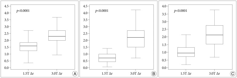
Fig. 3 A : The errors (Δx, Δy, Δz, and Δr) in the T1 sequence of the 1.5T and 3.0T MRIs. B : The errors in T2. C : The errors in fluid attenuated inversion recovery. All p-values are corrected by Bonferroni's method.

Fig. 4 The errors in the central area are shown in each MRI sequence. The difference between the 1.5T and 3.0T MRIs is statistically significant in all MRI sequences. All p-values are corrected by Bonferroni's method.

Fig. 5 The errors of all the axes are shown in each MRI sequence. The error in the x axis is the smallest regardless of the MRI sequence.
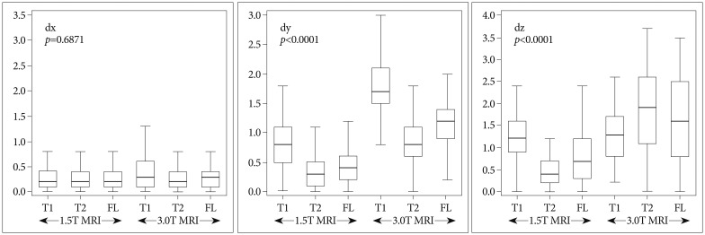
Fig. 6 The errors of all the MRI sequences are shown in each axis. The error in the T2 sequence is the smallest in the y and z axes. There is no difference in the x axis.
Cited by 1 articles
-
Avoiding a Collision in Gamma Knife Radiosurgery : A Modified Mask Fixation Method
Hyeong Cheol Moon, Doheui Lee, Byung Jun Min, Young Gyu Kim, Yun-Sik Dho
J Korean Neurosurg Soc. 2023;66(4):476-481. doi: 10.3340/jkns.2022.0164.
Reference
-
1. Andoh K, Nakamae H, Ohkoshi T, Odagiri K, Kyuma Y, Hayashi A. Technical note : enhanced MR-guided stereotaxic brain surgery with the patient under general anesthesia. AJNR Am J Neuroradiol. 1991; 12:135–138. PMID: 1899501.2. Bradford R, Thomas DG, Bydder GM. MRI-directed stereotactic biopsy of cerebral lesions. Acta Neurochir Suppl (Wien). 1987; 39:25–27. PMID: 3314381.
Article3. Choi DR, Ahn YC, Kim DY, Huh SJ, Lee JI. Accuracy in target localization in stereotactic radiosurgery. Med Dosim. 1997; 22:53–58. PMID: 9136109.
Article4. Dammann P, Kraff O, Wrede KH, Özkan N, Orzada S, Mueller OM, et al. Evaluation of hardware-related geometrical distortion in structural MRI at 7 Tesla for image-guided applications in neurosurgery. Acad Radiol. 2011; 18:910–916. PMID: 21549620.
Article5. Fransson A, Andreo P, Pötter R. Aspects of MR image distortions in radiotherapy treatment planning. Strahlenther Onkol. 2001; 177:59–73. PMID: 11233837.
Article6. Guo WY. Application of MR in stereotactic radiosurgery. J Magn Reson Imaging. 1998; 8:415–420. PMID: 9562069.7. Kangarlu A, Baertlein BA, Lee R, Ibrahim T, Yang L, Abduljalil AM, et al. Dielectric resonance phenomena in ultra high field MRI. J Comput Assist Tomogr. 1999; 23:821–831. PMID: 10589554.
Article8. Kondziolka D, Dempsey PK, Lunsford LD, Kestle JR, Dolan EJ, Kanal E, et al. A comparison between magnetic resonance imaging and computed tomography for stereotactic coordinate determination. Neurosurgery. 1992; 30:402–406. PMID: 1620305.
Article9. Loganathan AG, Chan MD, Alphonse N, Peiffer AM, Johnson AJ, McMullen KP, et al. Clinical outcomes of brain metastases treated with Gamma Knife radiosurgery with 3.0 T versus 1.5 T MRI-based treatment planning : have we finally optimised detection of occult brain metastases? J Med Imaging Radiat Oncol. 2012; 56:554–560. PMID: 23043576.
Article10. Lunsford LD, Martinez AJ, Latchaw RE. Stereotaxic surgery with a magnetic resonance- and computerized tomography-compatible system. J Neurosurg. 1986; 64:872–878. PMID: 3009738.
Article11. MacFadden D, Zhang B, Brock KK, Hodaie M, Laperriere N, Schwartz M, et al. Clinical evaluation of stereotactic target localization using 3-Tesla MRI for radiosurgery planning. Int J Radiat Oncol Biol Phys. 2010; 76:1472–1479. PMID: 19515512.
Article12. Mack A, Wolff R, Scheib S, Rieker M, Weltz D, Mack G, et al. Analyzing 3-tesla magnetic resonance imaging units for implementation in radiosurgery. J Neurosurg. 2005; 102 Suppl:158–164. PMID: 15662802.
Article13. Price RR, Axel L, Morgan T, Newman R, Perman W, Schneiders N, et al. Quality assurance methods and phantoms for magnetic resonance imaging : report of AAPM nuclear magnetic resonance Task Group No. 1. Med Phys. 1990; 17:287–295. PMID: 2333055.
Article14. Reinsberg SA, Doran SJ, Charles-Edwards EM, Leach MO. A complete distortion correction for MR images : II. Rectification of static-field inhomogeneities by similarity-based profile mapping. Phys Med Biol. 2005; 50:2651–2661. PMID: 15901960.
Article15. Tanner SF, Finnigan DJ, Khoo VS, Mayles P, Dearnaley DP, Leach MO. Radiotherapy planning of the pelvis using distortion corrected MR images : the removal of system distortions. Phys Med Biol. 2000; 45:2117–2132. PMID: 10958184.
Article16. Villemure JG, Marchand E, Peters T, Leroux G, Olivier A. Magnetic resonance imaging stereotaxy : recognition and utilization of the commissures. Appl Neurophysiol. 1987; 50:57–62. PMID: 3329884.
Article17. Watanabe Y, Lee CK, Gerbi BJ. Geometrical accuracy of a 3-tesla magnetic resonance imaging unit in Gamma Knife surgery. . J Neurosurg. 2006; 105 Suppl:190–193. PMID: 18503355.
Article
