Arthroscopic Analysis of the Radiologic Abnormalities of the Hip Associated with Anterior Femoroacetabular Impingement
- Affiliations
-
- 1Department of Orthopedic Surgery, Chungnam National University School of Medicine, Daejeon, Korea. dshwang@cnu.ac.kr
- 2Department of Orthopedic Surgery, Bumin Seoul Hospital, Seoul, Korea.
- 3Department of Orthopedic Surgery, College of Medicine, Konyang University, Daejeon, Korea.
- KMID: 2190771
- DOI: http://doi.org/10.5371/jkhs.2011.23.1.15
Abstract
- PURPOSE
We wanted to arthroscopically analyze the femoral osseous abnormalities (bumps) in hips with anterior femoroacetabular impingement (FAI) and the other radiologic abnormalities of the hip joint associated with this.
MATERIALS AND METHODS
We retrospectively reviewed the radiographs of 45 patients (51 hips) who underwent arthroscopic treatment under the impression of FAI from January to August, 2008. The indications for surgery included persistent hip pain, the absence of advanced osteoarthritis, physical examination or MRA findings consistent with an acetabualr labral tear. Preoperative and postoperative plain radiographs (pelvis AP, frog-leg lateral, cross table lateral and the false profile view) were taken. As the occasion demanded, 3D-CT or MRA were performed.
RESULTS
For the radiologic findings, a decreased head-neck offset (<8 mm) was seen in 15 hips. Femoral bumps were seen in 26 hips and among them, 11 hips were associated with acetabular retroversion, 5 hips were associated with a prominent acetabular rim and 13 hips were located on the flattening of the neck due to a decreased offset. Pistol grip deformity was found in 21 hips and acetabular retroversion was done in 32 hips. Regarding the secondary changes, spurs on the acetabulum of the femur were found in 14 hips and femoral bony cysts were found in 23 hips. Arthroscopically, all the hips had acetabular degenerative labral tear and the acetabular cartilage was injured in 32 hips (62.7%). Among them, 25 hips underwent acetabular retroversion.
CONCLUSION
Femoral osseous abnormalities are seen in various locations and these abnormalities have various shapes. A considerable number were associated with pincer impingement and they could produce a 'kissing lesion' between the acetabulum and femur. Identification of these abnormalities on radiographs aids confirming FAI in hips with symptomatic early osteoarthritis.
MeSH Terms
Figure
-
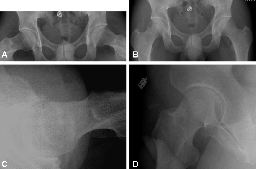
Fig. 1 Hip plain radiographies. (A) Hip AP. (B) Frog-leg lateral view. (C) Cross-table lateral view. (D) False profile view.
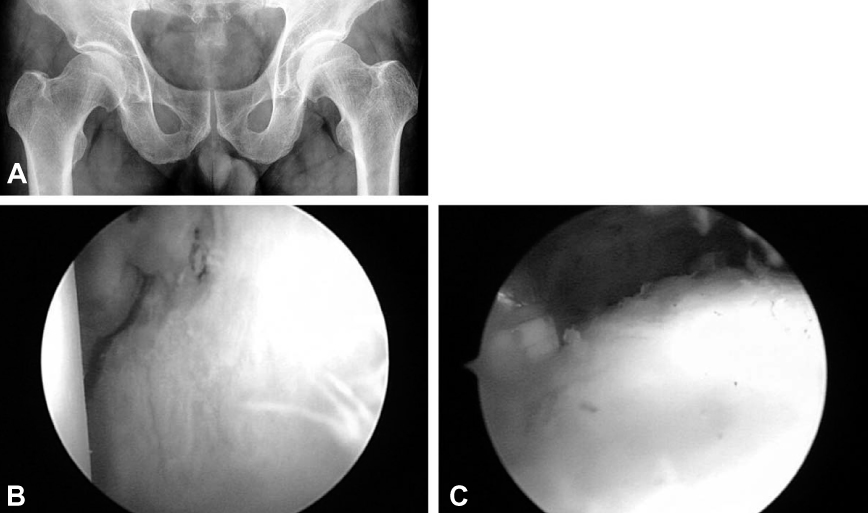
Fig. 2 Flattening of femoral neck with a retroversion. (A) Right hip: decreased curvature of femoral neck. (B) Acetabular labral tear. (C) Flattening of femoral neck.
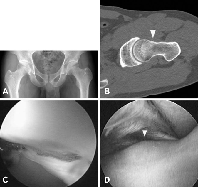
Fig. 3 Bump associated with acetabular retroversion. (A) Acetabular retroversion: Crossover sign (figure of eight) are positive on both hip joint. (B) CT: We found out bump formation (yellow arrow) on femoral neck. Arthroscopic findings: (C) Acetabular labral tear. (D) We could find out bump on femoral neck under arthroscopy.
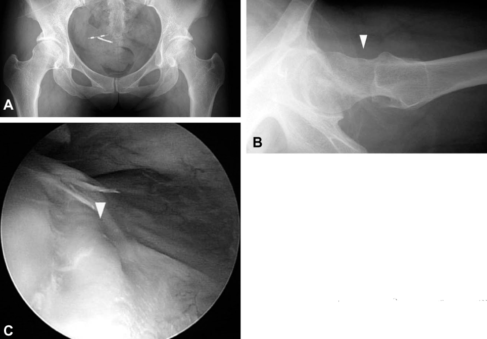
Fig. 4 Bump associated with prominent acetabular rim. (A, B) Both acetabula have a prominent acetabular rim and we can find bump (yellow arrow) on femoral neck at cross table lateral view. (C) Under arthroscopy, femoral bump (yellow arrow) was revealed on femoral neck consistent with prominent acetabular rim.
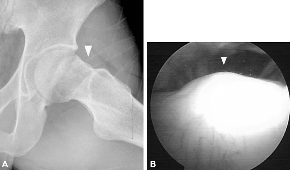
Fig. 5 Bump on flattening of femoral neck. (A) X-ray. (B) Arthroscopic finding.
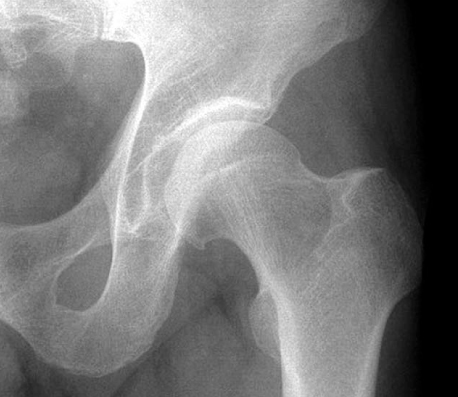
Fig. 6 Pistol grip deformity.
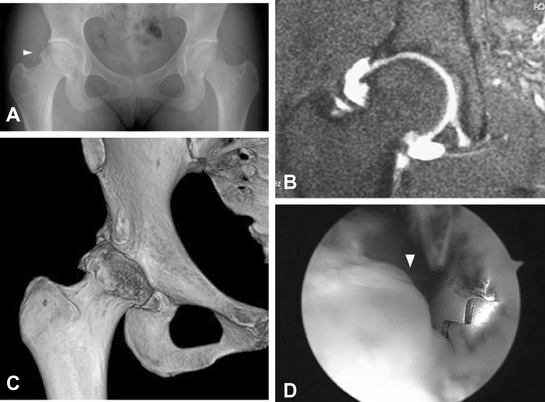
Fig. 7 Hook-shaped spur on head-neck junction. (A) Hook-shaped spur (yellow arrow) on femoral head-neck junction. (B) We check the hook on MR angiography. (C) 3 Dimensional CT image. (D) Arthroscopic image of spur (yellow arrow).
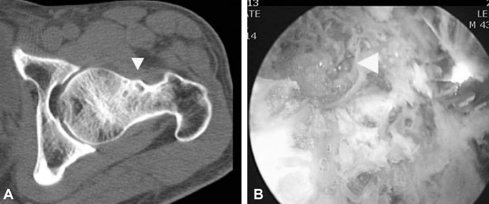
Fig. 8 Femoral bony cyst on femoral neck. (A) CT image: femoral bony cyst (yellow arrow) on femoral neck. (B) After bumpectomy, a hollow suggestive with cyst was revealed.
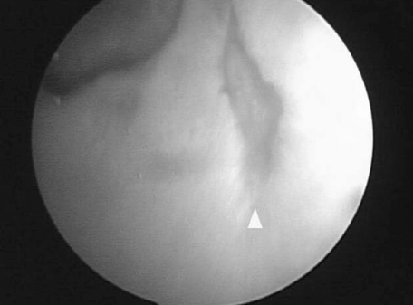
Fig. 9 Contrecoup lesion: arthroscopy showed a lesion (yellow arrow) on posteroinferior acetabular cartilage.
Reference
-
1. Leunig M, Ganz R. Femoroacetbular impingement. A common cause of hip complaints leading to arthrosis. Unfallchirurg. 2005. 108:9–10.2. McCarthy JC, Busconi B. The role of hip arthroscopy in the diagnosis and treatment of hip disease. Can J Surg. 1995. 38:Suppl 1. S13–S17.
Article3. McCarthy JC, Lee JA. Acetabular dysplasia: a paradigm of arthroscopic examination of chondral injuries. Clin Orthop Relat Res. 2002. 405:122–128.
Article4. McCarthy J, Noble P, Aluisio FV, Schuck M, Wright J, Lee JA. Anatomy, pathologic features, and treatment of acetabular labral tears. Clin Orthop Relat Res. 2003. 406:38–47.
Article5. Tönnis D, Heinecke A. Acetabular and femoral anteversion: relationship with osteoarthritis of the hip. J Bone Joint Surg Am. 1999. 81:1747–1770.6. Delaunay S, Dussault RG, Kaplan PA, Alford BA. Radiographic measurements of dysplastic adult hips. Skeletal Radiol. 1997. 26:75–81.
Article7. Ganz R, Parvizi J, Beck M, Leunig M, Nötzli H, Siebenrock KA. Femoroacetabular impingement: a cause for osteoarthritis of the hip. Clin Orthop Relat Res. 2003. 417:112–120.8. Lequesne M, Djian A. New radiologic angles for the study of the hip. The "false profile" of the pelvis. Vie Med. 1961. 42:1629–1641.9. Murphy SB, Ganz R, Müller ME. The prognosis in untreated dysplasia of the hip. A study of radiographic factors that predict the outcome. J Bone Joint Surg Am. 1995. 77:985–989.
Article10. Lequesne M, de Seze. False profile of the pelvis. A new radiographic incidence for the study of the hip. Its use in dysplasias and different coxopathices. Rev Rhum Mal Osteoartic. 1961. 28:643–652.11. Mast JW, Brunner RL, Zebrack J. Recognizing acetabular version in the radiographic presentation of hip dysplasia. Clin Orthop Relat Res. 2004. 418:48–53.
Article12. Reikerås O, Bjerkreim I, Kolbenstvedt A. Anteversion of the acetabulum and femoral neck in normals and in patients with osteoarthritis of the hip. Acta Orthop Scand. 1983. 54:18–23.
Article13. Reynolds D, Lucas J, Klaue K. Retroversion of the acetabulum. A cause of hip pain. J Bone Joint Surg Br. 1999. 81:281–288.14. Siebenrock KA, Schoeniger R, Ganz R. Anterior femoroacetabular impingement due to acetabular retroversion. Treatment with periacetabular osteotomy. J Bone Joint Surg Am. 2003. 85-A:278–286.15. Siebenrock KA, Wahab KH, Werlen S, Kalhor M, Leunig M, Ganz R. Abnormal extension of the femoral head epiphysis as a cause of cam impingement. Clin Orthop Relat Res. 2004. 418:54–60.
Article16. Eijer H, Myers SR, Ganz R. Anterior femoroacetabular impingement after femoral neck fractures. J Orthop Trauma. 2001. 15:475–481.
Article17. Tannast M, Siebenrock KA. Conventional radiographs to assess femoroacetabular impingement. Instr Course Lect. 2009. 58:203–212.18. Beck M, Leunig M, Parvizi J, Boutier V, Wyss D, Ganz R. Anterior femoroacetabular impingement: part II. Midterm results of surgical treatment. Clin Orthop Relat Res. 2004. 418:67–73.19. Giori NJ, Trousdale RT. Acetabular retroversion is associated with osteoarthritis of the hip. Clin Orthop Relat Res. 2003. 417:263–269.20. Millis MB, Kim YJ. Rationale of osteotomy and related procedures for hip preservation: a review. Clin Orthop Relat Res. 2002. 405:108–121.
Article21. Myers SR, Eijer H, Ganz R. Anterior femoroacetabular impingement after periacetabular osteotomy. Clin Orthop Relat Res. 1999. 363:93–99.
Article22. Kim YJ, Ganz R, Murphy SB, Buly RL, Millis MB. Hip joint-preserving surgery: beyond the classic osteotomy. Instr Course Lect. 2006. 55:145–158.23. Murray RO. The aetiology of primary osteoarthritis of the hip. Br J Radiol. 1965. 38:810–824.
Article24. Harris WH. Etiology of osteoarthritis of the hip. Clin Orthop Relat Res. 1986. 213:20–33.
Article25. Nötzli HP, Wyss TF, Stoecklin CH, Schmid MR, Treiber K, Hodler J. The contour of the femoral head-neck junction as a predictor for the risk of anterior impingement. J Bone Joint Surg Br. 2002. 84:556–560.
Article26. Dorrell JH, Catterall A. The torn acetabular labrum. J Bone Joint Surg Br. 1986. 68:400–403.
Article27. Eijer H, Leunig M, Mahomed MN, Ganz R. Cross-table lateral radiographs for screening of anterior femoral head-neck offset in patients with femoro-acetabular impingement. Hip Int. 2001. 11:37–41.
Article28. Ganz R, Gill TJ, Gautier E, Ganz K, Krügel N, Berlemann U. Surgical dislocation of the adult hip a technique with full access to the femoral head and acetabulum without the risk of avascular necrosis. J Bone Joint Surg Br. 2001. 83:1119–1124.29. Ito K, Minka MA 2nd, Leunig M, Werlen S, Ganz R. Femoroacetabular impingement and the cam-effect. A MRI-based, quantitative anatomical study of the femoral head-neck offset. J Bone Joint Surg Br. 2001. 83:171–176.30. Lavigne M, Parvizi J, Beck M, Siebenrock KA, Ganz R, Leunig M. Anterior femoroacetabular impingement: part I. Techniques of joint preserving surgery. Clin Orthop Relat Res. 2004. 418:61–66.31. Petersilge CA, Haque MA, Petersilge WJ, Lewin JS, Lieberman JM, Buly R. Acetabular labral tears: evaluation with MR arthrography. Radiology. 1996. 200:231–235.
Article32. Leunig M, Podeszwa D, Beck M, Werlen S, Ganz R. Magnetic resonance arthrography of labral disorders in hips with dysplasia and impingement. Clin Orthop Relat Res. 2004. 418:74–80.
Article33. Rab GT. The geometry of slipped capital femoral epiphysis: implications for movement, impingement, and corrective osteotomy. J Pediatr Orthop. 1999. 19:419–424.
Article34. Leunig M, Beck M, Kalhor M, Kim YJ, Werlen S, Ganz R. Fibrocystic changes at anterosuperior femoral neck: prevalence in hips with femoroacetabular impingement. Radiology. 2005. 236:237–246.
Article35. Philippon MJ, Dewing CB, Briggs KK. Kelly BT, Philippon MJ. Arthroscopic acetabular labral repair with rim trimming and femoral head-neck osteoplasty. 2010. New Jersy: SLACK;59–66.
- Full Text Links
-
- Actions
-
Cited
- CITED
-
- Close
- Share
-
- Similar articles
-
- Controversial Issues in Arthroscopic Surgery for Femoroacetabular Impingement
- Radiologic and Clinical Findings of Anterior Femoroacetabular Impingement in Early Osteoarthritis of the Hip
- Efficacy of Intra-articular Steroid Injection in Patients with Femoroacetabular Impingement
- Arthroscopic Treatment of Femoroacetabular Impingement of the Hip: 5-7 Years Result
- Arthroscopic Labral Repair Associated with Femoroacetabular Impingement: Short Term 2-5 Years Follow-up Results
