Analysis of crystalline structure of autogenous tooth bone graft material: X-Ray diffraction analysis
- Affiliations
-
- 1Department of Oral and Maxillofacial Surgery, College of Dentistry, Dankook University, Cheonan, Korea.
- 2Department of Prosthodontics, School of Dentistry and Dental Research Institute, Seoul National University, Seoul, Korea.
- 3Department of Oral and Maxillofacial Surgery, School of Dentistry, Chosun University, Gwangju, Korea.
- 4CTO, R&D Director, Korea Tooth Bank, Seoul, Korea.
- 5Department of Oral and Maxillofacial Surgery, Section of Dentistry, Seoul National University Bundang Hospital, School of Dentistry, Seoul National University, Seongnam, Korea. kyk0505@snubh.org
- KMID: 2189780
- DOI: http://doi.org/10.5125/jkaoms.2011.37.3.225
Abstract
- This study evaluated the mineral crystalline structure of an autogenous tooth bone graft material. The crystalline structures of the autogenous tooth bone graft material enamel (AutoBT E+), dentin (AutoBT D+), xenograft (BioOss), alloplastic material (MBCP), allograft (ICB) and autogenous mandibular cortical bone were compared using XRD. The XRD pattern of AutoBT dentin and ICB was similar to that of autogenous bone.
Keyword
MeSH Terms
Figure
-
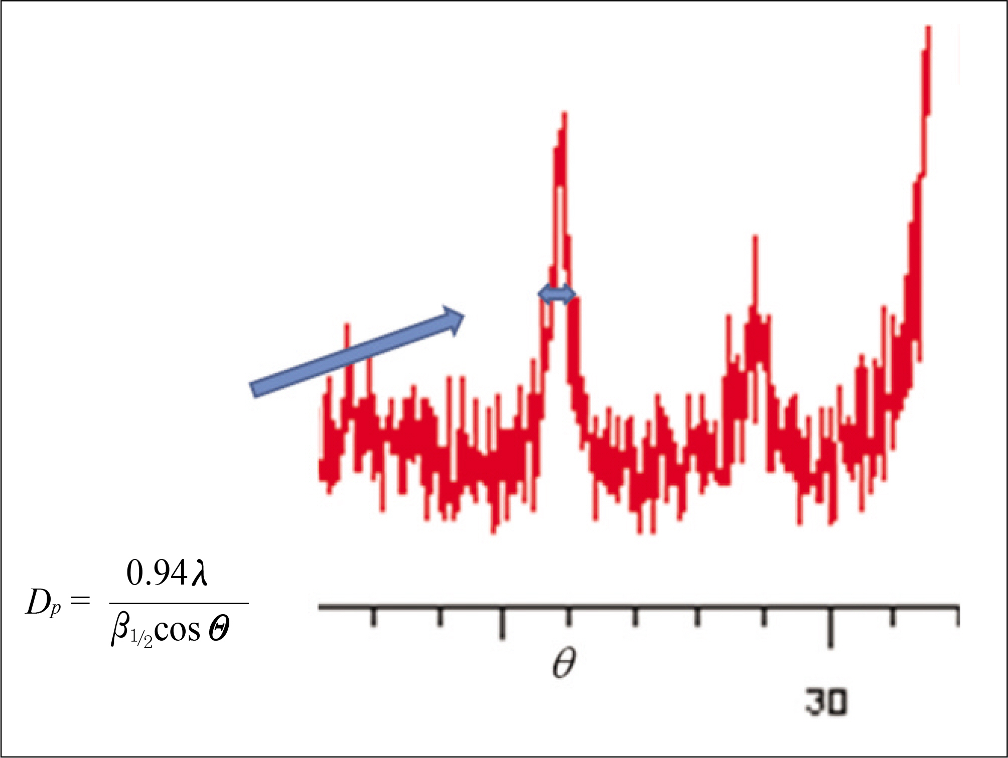
Fig. 1. Measurement of domain size. Arrow: width

Fig. 2. XRD patterns of a variety of bone graft materials. A: XRD pattern of autogenous bone, B: XRD pattern of AutoBT dentin, C: ARD pattern of AutoBT enamel, D: XRD pattern of ICB, E: XRD pattern of BioOss, F: XRD pattern of MBCP.(XRD: X-ray diffraction)
Cited by 4 articles
-
Bone graft material using teeth
Young-Kyun Kim
J Korean Assoc Oral Maxillofac Surg. 2012;38(3):134-138. doi: 10.5125/jkaoms.2012.38.3.134.Regenerative medicine for the reconstruction of hard tissue defects in oral and maxillofacial surgery
Young-Kyun Kim
J Korean Assoc Oral Maxillofac Surg. 2012;38(2):69-70. doi: 10.5125/jkaoms.2012.38.2.69.Comparison of autogenous tooth bone graft and synthetic bone graft materials used for bone resorption around implants after crestal approach sinus lifting: a retrospective study
Young-Kyun Kim, Junho Lee, Ji-Young Yun, Pil-Young Yun, In-Woong Um
J Periodontal Implant Sci. 2014;44(5):216-221. doi: 10.5051/jpis.2014.44.5.216.Tooth-derived bone graft material
Young-Kyun Kim, Junho Lee, In-Woong Um, Kyung-Wook Kim, Masaru Murata, Toshiyuki Akazawa, Masaharu Mitsugi
J Korean Assoc Oral Maxillofac Surg. 2013;39(3):103-111. doi: 10.5125/jkaoms.2013.39.3.103.
Reference
-
References
1. Kim YK, Yeo HH, Ryu CH, Lee HB, Byun UR, Cho JO. An experimental study on the tissue reaction of toothash implanted in mandible body of the mature dog. J Korean Assoc Maxillofac Plast Reconstr Surg. 1993; 15:129–36.2. Kim YK, Yeo HH, Yang IS, Seo JH, Cho JO. Implantation of toothash combined with plaster of paris: experimental study. J Korean Assoc Maxillofac Plast Reconstr Surg. 1994; 16:122–9.3. Kim YK, Kim SG, Byeon JH, Lee HJ, Um IU, Lim SC, et al. Development of a novel bone grafting material using autogenous teeth. Oral Surg Oral Med Oral Pathol Oral Radiol Endod. 2010; 109:496–503.
Article4. Kim YK, Lee HJ, Kim SG, Um IW, Lim SC, Kim SY. Analysis of inorganic component and SEM analysis of autogenous teeth bone graft material and histomorphometric analysis after graft. J Korean Acad Implant Dent. 2009; 28:1–9.5. Kim YK, Lee JY. The evaluation of postoperative safety of autogenous teeth bone graft. J Korean Acad Implant Dent. 2009; 28:29–35.6. Klug HP, Alexander LE. X-ray diffraction procedures for polycrystalline and amorphous materials. 2nd ed.New York: Wiley-Interscience;1974.7. Kim YK, Kim SG, Lee BG. Bone graft and implant: vol 1. Bone biology and bone graft material. Seoul: Narae Pub Co.;2007. p. 171–224.8. Tadic D, Epple M. A thorough physicochemical characterisation of 14 calcium phosphate-based bone substitution materials in comparison to natural bone. Biomaterials. 2004; 25:987–94.
Article9. Kirik SD, Solovyov LA, Blokhin AI, Yakimov IS. Structures of. Acta Crystallogr B. 2000; 56:419–25.10. Balasundaram G, Sato M, Webster TJ. Using hydroxyapatite nanoparticles and decreased crystallinity to promote osteoblast adhesion similar to functionalizing with RGD. Biomaterils. 2006; 27:2798–805.
Article11. Kim YK, Kim SG, Jin SC, Son JS, Kim SY, Um IW. Analysis of the Inorganic Component of Autogenous Tooth Bone Graft Material. J Nanosci Nanotechnol 2011 accepted.12. Glimcher MJ. Molecular biology of mineralized tissues with particular reference to bone. Rev Mod Phys. 1959; 31:359–93.
Article13. Lee SH. Low Crystalline hydroxyl carbonate apatite. J Korean Dental Assoc. 2006; 44:524–33.
- Full Text Links
-
- Actions
-
Cited
- CITED
-
- Close
- Share
-
- Similar articles
-
- Bone graft material using teeth
- Clinical Study on the Alveolar Bone Repair Capacity of Dentin Matrix Block
- Ridge Augmentation Using Block Type of Autogenous Tooth Bone Graft Material in Severe Alveolar Bone Resorption of Single Tooth: Case Report
- Evaluation of the Healing Process of Autogenous Tooth Bone Graft Material Nine Months after Sinus Bone Graft: Micromorphometric and Histological Evaluation
- Clinical Study on the Efficacy of the Autogenous Tooth Bone Graft Material (AutoBT)
