Expansile dentigerous cyst invading the entire maxillary sinus: a case report
- Affiliations
-
- 1Department of Dentistry, Dongsan Medical Center, Keimyung University, Daegu, Korea. nkyp@dsmc.or.kr
- KMID: 2189632
- DOI: http://doi.org/10.5125/jkaoms.2012.38.4.245
Abstract
- Reported cases of a large dentigerous cyst involving the whole maxillary sinus are uncommon. A 22-year-old female patient suffering from swelling of the right infraorbital area and cheek with dull pain was referred to our department. Findings on computed tomography (CT) and magnetic resonance imaging (MRI) revealed a huge mass containing a displaced maxillary third molar involving the right maxillary sinus as a whole, with partial erosion of the posterior sinus cortical bone. Under general anesthesia, the mass was enucleated using the Caldwell-Luc approach, and, following histopathological analysis, was diagnosed as a dentigerous cyst. The case was followed for a period of seven years, and no evidence of sinus infection or recurring cyst formation was observed during that time.
MeSH Terms
Figure
-
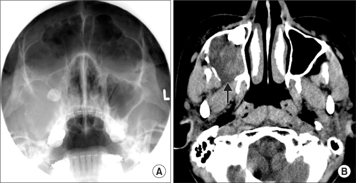
Fig. 1 A. Pre-operative Water's view; haziness of right maxillary sinus with thinned lateral wall and dislocated maxillary third molar were detected. B. Partially disrupted posterior sinus wall with irregular margin (arrow) on axial computed tomography image.
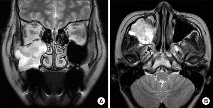
Fig. 2 A. Pre-operative frontal magnetic resonance (MR) image. B. Hypointense dark rim (arrow) is seen at posterior border of the mass in axial MR image.
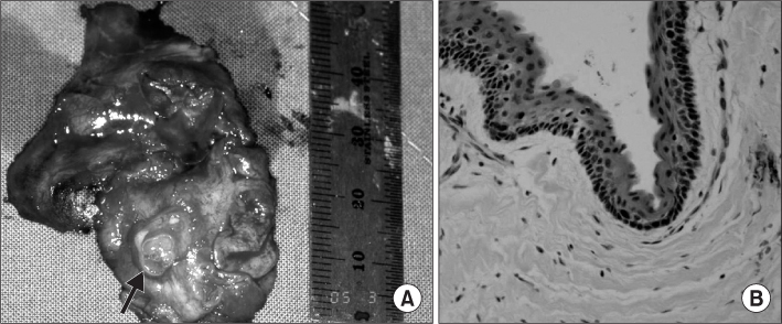
Fig. 3 A. The excised mass with a displaced third molar contained (arrow). B. Histopathologically, nonkeratinizing squamous epithelial lining, cholesterol clefts and Rushton body are seen (H&E staining, ×200).

Fig. 4 Post-operative panoramic view after 7 years.
Cited by 3 articles
-
Expansile keratocystic odontogenic tumor in the maxilla: immunohistochemical studies and review of literature
June-Ho Byun, Young-Hoon Kang, Mun-Jeong Choi, Bong-Wook Park
J Korean Assoc Oral Maxillofac Surg. 2013;39(4):182-187. doi: 10.5125/jkaoms.2013.39.4.182.Management of rare ectopic teeth eruption: case series
Olutayo James, Ibrahim Kayode Suleiman, Mukhtar Modibbo Ahmad, Hector Oladapo Olasoji
J Korean Assoc Oral Maxillofac Surg. 2023;49(2):86-90. doi: 10.5125/jkaoms.2023.49.2.86.Is conservative treatment (enucleation using modified Carnoy’s solution) of odontogenic keratocyst in the maxilla good prognosis?
Woo Young Jeon, Jung Ho Park, Jeong-Kui Ku, Jin-A Baek, Seung-O Ko
J Korean Assoc Oral Maxillofac Surg. 2023;49(5):287-291. doi: 10.5125/jkaoms.2023.49.5.287.
Reference
-
1. Kim SG, Park CY, Kang TH, Jang HS. Clinicopathologic study on cysts and postoperative cyst in maxillary sinus. J Korean Assoc Maxillofac Plast Reconstr Surg. 2000. 22:568–576.2. Altas E, Karasen RM, Yilmaz AB, Aktan B, Kocer I, Erman Z. A case of a large dentigerous cyst containing a canine tooth in the maxillary antrum leading to epiphora. J Laryngol Otol. 1997. 111:641–643.
Article3. Motamedi MH, Talesh KT. Management of extensive dentigerous cysts. Br Dent J. 2005. 198:203–206.
Article4. Kim KW, Lee JH. Clinical study of cysts in the jaws. J Korean Assoc Maxillofac Plast Reconstr Surg. 1999. 21:166–173.5. Park TW. Clinico-radiological study of cyst of the jaw. J Korean Acad Oral Maxillofac Radiol. 1983. 13:163–168.6. Hong SP, Lee JI, Shin HI, Choi HR, Kim EC, Park HR. Contemporary oral and maxillofacial pathology. 1999. Seoul: Jeong Won Publishing Co..7. Im CY. Color atlas of oral pathology. 1992. Seoul: Korea Medical Publishing Co..8. Albright CR, Hennig GH. Large dentigerous cyst of the maxilla near the maxillary sinus: report of case. J Am Dent Assoc. 1971. 83:1112–1115.
Article9. Avitia S, Hamilton JS, Osborne RF. Dentigerous cyst presenting as orbital proptosis. Ear Nose Throat J. 2007. 86:23–24.
Article10. Na CY, Choi KS. A radiographic study of differential diagnosis between dentigerous cysts and unicystic ameloblastomas. J Korean Acad Oral Maxillofac Radiol. 1993. 23:255–264.11. Weber AL. Imaging of cysts and odontogenic tumors of the jaw. Definition and classification. Radiol Clin North Am. 1993. 31:101–120.12. Robinson L, Martinez MG. Unicystic ameloblastoma: a prognostically distinct entity. Cancer. 1977. 40:2278–2285.
Article13. Som PM, Dillon WP, Curtin HD, Fullerton GD, Lidov M. Hypointense paranasal sinus foci: differential diagnosis with MR imaging and relation to CT findings. Radiology. 1990. 176:777–781.
Article14. Dagistan S, Cakur B, Goregen M. A dentigerous cyst containing an ectopic canine tooth below the floor of the maxillary sinus: a case report. J Oral Sci. 2007. 49:249–252.
Article15. Chuong R. Dentigerous cyst involving maxillary sinus: report of case. J Am Dent Assoc. 1984. 109:59–60.
Article16. Hyomoto M, Kawakami M, Inoue M, Kirita T. Clinical conditions for eruption of maxillary canines and mandibular premolars associated with dentigerous cysts. Am J Orthod Dentofacial Orthop. 2003. 124:515–520.
Article17. Ertas U, Yavuz MS. Interesting eruption of 4 teeth associated with a large dentigerous cyst in mandible by only marsupialization. J Oral Maxillofac Surg. 2003. 61:728–730.
Article
- Full Text Links
-
- Actions
-
Cited
- CITED
-
- Close
- Share
-
- Similar articles
-
- Endonasal Removal of Dentigerous Cyst in the Maxillary Sinus
- Huge Dentigerous Cyst
- A Long-term Follow-Up Case of Enucleation of Dentigerous Cyst in the Maxilla: Case Report
- Endoscopic approach for treatment of dentigerous cyst in maxillary sinus
- An Inflammatory Dentigerous Cyst Shows Rim Uptake on Bone Scan: A Case Report
