Correlation between glucose transporter type-1 expression and 18F-FDG uptake on PET in oral cancer
- Affiliations
-
- 1Department of Oral and Maxillofacial Surgery, College of Dentistry, Dankook University, Cheonan, Korea. kimchoms@dankook.ac.kr
- KMID: 2189628
- DOI: http://doi.org/10.5125/jkaoms.2012.38.4.212
Abstract
OBJECTIVES
Fluorine-18 fluorodeoxyglucose positron emission tomography (18F-FDG PET) is a non-invasive diagnostic tool for many human cancers wherein glucose uptake transporter-1 (GLUT-1) acts as a main transporter in the uptake of 18F-FDG in cancer cells. Increased expression of glucose transporter-1 has been reported in many human cancers. In this study, we investigated the correlation between 18F-FDG accumulation and expression of GLUT-1 in oral cancer.
MATERIALS AND METHODS
We evaluated 42 patients diagnosed with oral squamous cell carcinoma (OSCC) and malignant salivary gland tumor as confirmed by histology. 42 patients underwent pre-operative 18F-FDG PET, with the maximum standardized uptake value (SUVmax) measured in each case. Immunohistochemical staining was done for each histological specimen, and results were evaluated post-operatively according to the percentage (%) of positive area, intensity, and staining score.
RESULTS
For OSCC, SUVmax significantly increased as T stage of tumor classification increased. For malignant salivary gland tumor, SUVmax significantly increased as T stage of tumor classification increased. For OSCC, GLUT-1 was expressed in all 36 cases. GLUT-1 staining score (GSS) increased as T stage of tumor classification increased, with the difference statistically significant. For malignant salivary gland tumor, GLUT-1 expression was observed in all 6 cases; average GSS was significantly higher in patients with cervical lymph node metastasis than that in patients without cervical lymph node metastasis. Average GSS was higher in OSCC (11.11+/-1.75) than in malignant salivary gland tumor (5.33+/-3.50). No statistically significant correlation between GSS and SUVmax was observed in OSCC or in malignant salivary gland tumor.
CONCLUSION
We found no statistically significant correlation between GSS and SUVmax in OSCC or in malignant salivary gland tumor. Studies on the various uses of GLUT during 18F-FDG uptake and SUV and GLUT as tumor prognosis factor need to be conducted through further investigation with large samples.
Keyword
MeSH Terms
Figure
-
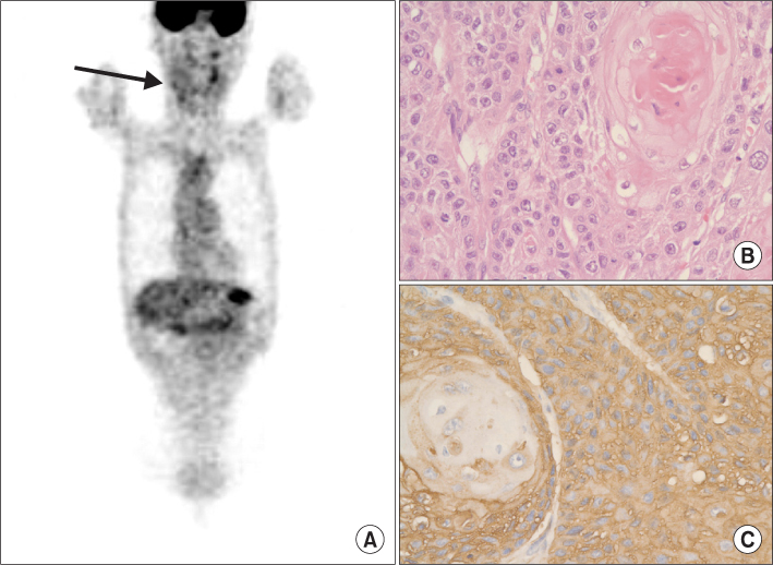
Fig. 1 Well-differentiated squamous cell carcinoma in the oral floor of a 73-year-old man (case No. 2). A. Positron emission tomography. Coronal maximum intensity projection image with increased fluorodeoxyglucose accumulation in the right floor of mouth (arrow). B. Well-differentiated squamous cell carcinoma confirmed on H&E staining (×400). C. Immunohistochemical staining score for glucose uptake transporter-1 in tumor cells was 12 (strong intensity and more than 75% of positive area) (×400).

Fig. 2 Mucoepidermoid carcinoma in the right maxilla of a 43-year-old woman (case No. 37). A. Positron emission tomography (PET). Coronal maximum intensity projection image with increased fluorodeoxyglucose accumulation in the right floor of mouth (arrow). B. Fused PET and computed tomography images. A tumor shows maximum standardized uptake value of 3.2. C. Mucoepidermoid carcinoma confirmed on H&E staining (×200). D. Immunohistochemical staining score for glucose uptake transporter-1 in tumor cells was 4 (moderate intensity and 25-50% of positive area) (×200).

Fig. 3 Adenoid cystic carcinoma in the right maxilla of a 78-year-old woman (case No. 39). A. Positron emission tomography (PET). Coronal maximum intensity projection image with increased fluorodeoxyglucose accumulation in the right floor of mouth (arrow). B. Fused PET and computed tomography images. A tumor shows maximum standardized uptake value of 5.2. C. Adenoid cystic carcinoma confirmed on H&E staining (×200). D. Immunohistochemical staining score for glucose uptake transporter-1 in tumor cells was 2 (weak intensity and 25-50% of positive area) (×200).
Reference
-
1. Vokes EE, Weichselbaum RR, Lippman SM, Hong WK. Head and neck cancer. N Engl J Med. 1993. 328:184–194.
Article2. Jemal A, Siegel R, Ward E, Murray T, Xu J, Thun MJ. Cancer statistics, 2007. CA Cancer J Clin. 2007. 57:43–66.
Article3. Oehr P, Ruhlmann J, Biersack HJ. FDG-PET in clinical oncology: review of the literature and report of one institution's experience. J Investig Med. 1999. 47:452–461.4. Barrington SF. Atlas of clinical positron emission tomography. 2006. 2nd ed. London: Hodder Arnold;25–120.5. Kayano T, Burant CF, Fukumoto H, Gould GW, Fan YS, Eddy RL, et al. Human facilitative glucose transporters. Isolation, functional characterization, and gene localization of cDNAs encoding an isoform (GLUT5) expressed in small intestine, kidney, muscle, and adipose tissue and an unusual glucose transporter pseudogene-like sequence (GLUT6). J Biol Chem. 1990. 265:13276–13282.
Article6. Chung JK, Lee YJ, Kim C, Choi SR, Kim M, Lee K, et al. Mechanisms related to [18F]fluorodeoxyglucose uptake of human colon cancers transplanted in nude mice. J Nucl Med. 1999. 40:339–346.7. Higashi T, Saga T, Nakamoto Y, Ishimori T, Mamede MH, Wada M, et al. Relationship between retention index in dual-phase (18)F-FDG PET, and hexokinase-II and glucose transporter-1 expression in pancreatic cancer. J Nucl Med. 2002. 43:173–180.8. Chung JK, Lee YJ, Kim SK, Jeong JM, Lee DS, Lee MC. Comparison of [18F]fluorodeoxyglucose uptake with glucose transporter-1 expression and proliferation rate in human glioma and non-small-cell lung cancer. Nucl Med Commun. 2004. 25:11–17.
Article9. Higashi K, Ueda Y, Sakurai A, Wang XM, Xu L, Murakami M, et al. Correlation of Glut-1 glucose transporter expression with. Eur J Nucl Med. 2000. 27:1778–1785.
Article10. Schönberger J, Rüschoff J, Grimm D, Marienhagen J, Rümmele P, Meyringer R, et al. Glucose transporter 1 gene expression is related to thyroid neoplasms with an unfavorable prognosis: an immunohistochemical study. Thyroid. 2002. 12:747–754.
Article11. Lee BC, Shim YS, Lee YS, Lee GH, Seong NY, Hong SC, et al. SUV analysis of PET scan for prognostic factor of head and neck Cancer. Korean J Otolaryngol-Head Neck Surg. 2003. 46:955–958.12. Kim TY, Kim WB, Ryu JS, Gong G, Hong SJ, Shong YK. 18F-fluorodeoxyglucose uptake in thyroid from positron emission tomogram (PET) for evaluation in cancer patients: high prevalence of malignancy in thyroid PET incidentaloma. Laryngoscope. 2005. 115:1074–1078.
Article13. Minn H, Lapela M, Klemi PJ, Grénman R, Leskinen S, Lindholm P, et al. Prediction of survival with fluorine-18-fluoro-deoxyglucose and PET in head and neck cancer. J Nucl Med. 1997. 38:1907–1911.14. Torizuka T, Tanizaki Y, Kanno T, Futatsubashi M, Naitou K, Ueda Y, et al. Prognostic value of 18F-FDG PET in patients with head and neck squamous cell cancer. AJR Am J Roentgenol. 2009. 192:W156–W160.15. Kunkel M, Reichert TE, Benz P, Lehr HA, Jeong JH, Wieand S, et al. Overexpression of Glut-1 and increased glucose metabolism in tumors are associated with a poor prognosis in patients with oral squamous cell carcinoma. Cancer. 2003. 97:1015–1024.
Article16. Vansteenkiste JF, Stroobants SG, Dupont PJ, De Leyn PR, Verbeken EK, Deneffe GJ, et al. Leuven Lung Cancer Group. Prognostic importance of the standardized uptake value on (18)F-fluoro-2-deoxy-glucose-positron emission tomography scan in non-small-cell lung cancer: An analysis of 125 cases. J Clin Oncol. 1999. 17:3201–3206.
Article17. Kitagawa Y, Sadato N, Azuma H, Ogasawara T, Yoshida M, Ishii Y, et al. FDG PET to evaluate combined intra-arterial chemotherapy and radiotherapy of head and neck neoplasms. J Nucl Med. 1999. 40:1132–1137.18. Tateishi U, Yamaguchi U, Seki K, Terauchi T, Arai Y, Hasegawa T. Glut-1 expression and enhanced glucose metabolism are associated with tumour grade in bone and soft tissue sarcomas: a prospective evaluation by [18F]fluorodeoxyglucose positron emission tomography. Eur J Nucl Med Mol Imaging. 2006. 33:683–691.
Article19. Mochiki E, Kuwano H, Katoh H, Asao T, Oriuchi N, Endo K. Evaluation of 18F-2-deoxy-2-fluoro-D-glucose positron emission tomography for gastric cancer. World J Surg. 2004. 28:247–253.
Article20. Yen TC, See LC, Lai CH, Yah-Huei CW, Ng KK, Ma SY, et al. 18F-FDG uptake in squamous cell carcinoma of the cervix is correlated with glucose transporter 1 expression. J Nucl Med. 2004. 45:22–29.21. Kurokawa T, Yoshida Y, Kawahara K, Tsuchida T, Okazawa H, Fujibayashi Y, et al. Expression of GLUT-1 glucose transfer, cellular proliferation activity and grade of tumor correlate with [F-18]-fluorodeoxyglucose uptake by positron emission tomography in epithelial tumors of the ovary. Int J Cancer. 2004. 109:926–932.
Article22. Tohma T, Okazumi S, Makino H, Cho A, Mochiduki R, Shuto K, et al. Relationship between glucose transporter, hexokinase and FDG-PET in esophageal cancer. Hepatogastroenterology. 2005. 52:486–490.23. Yasuda M, Ogane N, Hayashi H, Kameda Y, Miyagi Y, Iida T, et al. Glucose transporter-1 expression in the thyroid gland: clinicopathological significance for papillary carcinoma. Oncol Rep. 2005. 14:1499–1504.
Article24. Mantych GJ, James DE, Chung HD, Devaskar SU. Cellular localization and characterization of Glut 3 glucose transporter isoform in human brain. Endocrinology. 1992. 131:1270–1278.
Article25. Zhou S, Wang S, Wu Q, Fan J, Wang Q. Expression of glucose transporter-1 and -3 in the head and neck carcinoma--the correlation of the expression with the biological behaviors. ORL J Otorhinolaryngol Relat Spec. 2008. 70:189–194.
Article26. Younes M, Lechago LV, Lechago J. Overexpression of the human erythrocyte glucose transporter occurs as a late event in human colorectal carcinogenesis and is associated with an increased incidence of lymph node metastases. Clin Cancer Res. 1996. 2:1151–1154.27. Tian M, Zhang H, Nakasone Y, Mogi K, Endo K. Expression of Glut-1 and Glut-3 in untreated oral squamous cell carcinoma compared with FDG accumulation in a PET study. Eur J Nucl Med Mol Imaging. 2004. 31:5–12.
Article28. Gu J, Yamamoto H, Fukunaga H, Danno K, Takemasa I, Ikeda M, et al. Correlation of GLUT-1 overexpression, tumor size, and depth of invasion with 18F-2-fluoro-2-deoxy-D-glucose uptake by positron emission tomography in colorectal cancer. Dig Dis Sci. 2006. 51:2198–2205.
Article29. Rege S, Safa AA, Chaiken L, Hoh C, Juillard G, Withers HR. Positron emission tomography: an independent indicator of radiocurability in head and neck carcinomas. Am J Clin Oncol. 2000. 23:164–169.
Article
- Full Text Links
-
- Actions
-
Cited
- CITED
-
- Close
- Share
-
- Similar articles
-
- PET oncology; Lung cancer
- Characteristics of [ 18F ] fluorodeoxyglucose Uptake in Human Colon Cancer Cells
- Correlation of Glucose Transporter-1 Expression With Uptake of 18F-Fluorodeoxyglucose Positron Emission Tomography in Thyroid Papillary Carcinoma
- Glut1 Expression and FDG Uptake in Non-small Cell Lung Carcinoma: Its Relationship to Histopathologic Types and Proliferation Rate
- Correlation of GLUT-1 Expression and F-18-FDG Uptake on Positron Emission Tomography in Breast Carcinoma
