J Korean Assoc Oral Maxillofac Surg.
2016 Feb;42(1):43-46. 10.5125/jkaoms.2016.42.1.43.
Ameloblastic carcinoma of the maxilla: a report of two cases and a review of the literature
- Affiliations
-
- 1Maxillofacial Unit, Ahmadu Bello University Teaching Hospital, Zaria, Nigeria. benfometey@hotmail.com
- 2Army Dental Centre, Military Hospital, Lagos, Nigeria.
- 3Department of Medical Laboratory, 44 Nigerian Army Reference Hospital, Kaduna, Nigeria.
- KMID: 2189412
- DOI: http://doi.org/10.5125/jkaoms.2016.42.1.43
Abstract
- Ameloblastic carcinoma is a malignant form of ameloblastoma defined by histological evidence of malignancy in primary, recurrent, or metastatic tumor. Such a tumor is rare, and the maxilla is an unusual site. Due to its rarity, the characteristics of this tumor in the maxilla have not been well described. Case 1: A 55-year-old, ill-appearing Nigerian male presented to our center with left maxillary swelling of seven-year duration. The swelling had been slow-growing and painless until one year prior, when the growth became rapid and was coupled with severe pain. The swelling affected both oral function and facial esthetics, and the patient reported difficulty breathing. There was a maxillary, ulcerated swelling extending from teeth 12 to 18 and blocking the left nostril. The involved teeth were moderately mobile. Case 2: A 32-year-old male farmer presented with recurrent right maxillary swelling of six-year duration. Prior to this episode, he had undergone surgery for ameloblastoma (follicular type). The present swelling was fungating through the skin and protruding into the right nostril. Ameloblastic carcinoma is an aggressive odontogenic tumor that requires aggressive surgical treatment.
Keyword
MeSH Terms
Figure
-
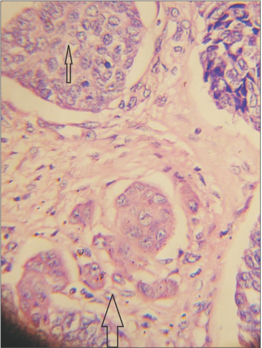
Fig. 1 Ameloblastic carcinoma shows extensive follicular basaloid (H&E staining, ×400). Arrows indicate the transformed malignant area showing basaloid malignant cells with focal stromal invasion.
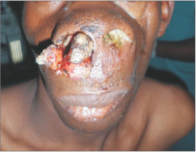
Fig. 2 Case 2, with a lesion protruding from the nose.

Fig. 3 Intraoperative photograph.

Fig. 4 Postoperative photograph.
Reference
-
1. Small LA, Waldron CA. Ameloblastoma of the jaws. Oral Surg Oral Med Oral Pathol. 1955; 8:281–297. PMID: 14356722.2. Jackson IT, Callan PP, Forté RA. An anatomical classification of maxillary ameloblastoma as an aid to surgical treatment. J Craniomaxillofac Surg. 1996; 24:230–236. PMID: 8880449.
Article3. Ajagbe HA, Daramola JO. Ameloblastoma: a survey of 199 cases in the University of College Hospital, Ibadan, Nigeria. J Natl Med Assoc. 1987; 79:324–327. PMID: 3573061.4. Horváth A, Horváth E, Popşor S. Mandibular ameloblastic carcinoma in a young patient. Rom J Morphol Embryol. 2012; 53:179–183. PMID: 22395519.5. Pindborg JJ, Kramer IR, Torloni H. Histological typing of odontogenic tumors, jaw cysts and allied lesions. Berlin: Springer-Verlag;1972. p. 35–36.6. Shafer WG, Hine MK, Levy BM. A textbook of oral pathology. 4th ed. Philadelphia: WB Saunders;1983. p. 280–281.7. Barnes L, Eveson JW, Reichart P, Sidransky D. World Health Organization Classification of Tumours: pathology and genetics of head and neck tumours. Lyon: IARC Press;2005. p. 283–328.8. Ward BB, Edlund S, Sciubba J, Helman JI. Ameloblastic carcinoma (primary type) isolated to the anterior maxilla: case report with review of the literature. J Oral Maxillofac Surg. 2007; 65:1800–1803. PMID: 17719401.
Article9. Dhir K, Sciubba J, Tufano RP. Ameloblastic carcinoma of the maxilla. Oral Oncol. 2003; 39:736–741. PMID: 12907214.
Article10. Kruse AL, Zwahlen RA, Grätz KW. New classification of maxillary ameloblastic carcinoma based on an evidence-based literature review over the last 60 years. Head Neck Oncol. 2009; 1:31. PMID: 19674470.
Article11. Adebayo ET, Ajike SO, Adekeye EO. A review of 318 odontogenic tumors in Kaduna, Nigeria. J Oral Maxillofac Surg. 2005; 63:811–819. PMID: 15944979.
Article12. Regezi JA, Kerr DA, Courtney RM. Odontogenic tumors: analysis of 706 cases. J Oral Surg. 1978; 36:771–778. PMID: 280645.13. Corio RL, Goldblatt LI, Edwards PA, Hartman KS. Ameloblastic carcinoma: a clinicopathologic study and assessment of eight cases. Oral Surg Oral Med Oral Pathol. 1987; 64:570–576. PMID: 3313152.
Article14. Ramesh M, Sekar B, Murali S, Mathew S, Chacko J, Paul G. Ameloblastic carcinoma: review and histopathology of 5 cases. Oral Maxillofac Pathol J. 2011; 2:154–160.15. Angiero F, Borloni R, Macchi M, Stefani M. Ameloblastic carcinoma of the maxillary sinus. Anticancer Res. 2008; 28:3847–3854. PMID: 19192639.16. Koul R, Binahmed A, Dubey A, Nason R, Cooke AL. Maxillary ameloblastic carcinoma. J Hong Kong Coll Radiol. 2008; 11:32–34.17. Marx RE, Stern D, et al. Oral and maxillofacial pathology: a rationale for diagnosis and treatment. Chicago: Quintessence Publishing;2003. p. 657.18. Philip M, Morris CG, Werning JW, Mendenhall WM. Radiotherapy in the treatment of ameloblastic carcinoma. J Hong Kong Coll Radiol. 2005; 8:157–161.
