Severe Genu Recurvatum after a Closing-wedge High Tibial Osteotomy: A Case Report
- Affiliations
-
- 1Department of Orthopaedic Surgery, Pusan National University College of Medicine, Pusan, Korea. jtsuh@pusan.ac.kr
- KMID: 2186458
- DOI: http://doi.org/10.4055/jkoa.2008.43.3.391
Abstract
- Although a closing wedge high tibial osteotomy can decrease the posterior tibial slope, there are no reports of severe genu recurvatum after a closing wedge osteotomy that required corrective surgery. A 54-year-old male with medial compartment osteoarthritis and genu varum was treated with a closing wedge high tibial osteotomy in another hospital, which led to severe recurvatum because of surgical failure. He complained of knee pain and a severe deformity, but of which were corrected by an open wedge corrective osteotomy, a two-wedge bicortical autograft reconstruction, and double plate fixation. Surgeons should pay close attention to both varus deformity correction and changes in the posterior tibial slope during a closing wedge high tibial osteotomy.
Figure
-
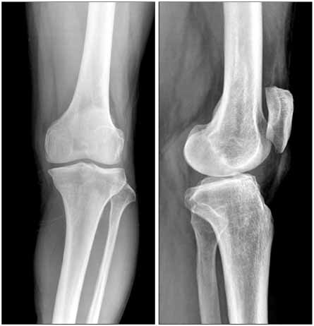
Fig. 1 Preoperative radiographs of a 54-year-old male showing genu varum with medial compartment osteoarthritis. The femorotibial angle was varus 5 degree and the tibial posterior slope was 7 degrees.
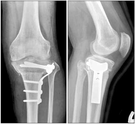
Fig. 2 Three months after the closing wedge osteotomy in another hospital, postoperative radiographs show the correction of genu varum but severe genu recurvatum. The femorotibial angle was valgus 2 degrees and the tibial posterior slope was -15 degrees.
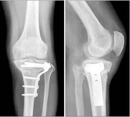
Fig. 3 Ten months after the closing wedge osteotomy, radiographs show persistent genu recurvatum and loss of valgus correction. The femorotibial angle was varus 2 degrees and the tibial posterior slope was -14 degrees.
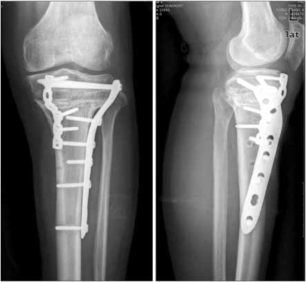
Fig. 4 Four months after the corrective osteotomy, radiographs show the correction of genu recurvatum and genu varum. The femorotibial angle was valgus 5 degrees and the tibial posterior slope was 4 degrees.
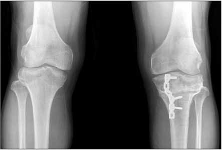
Fig. 5 A standing AP radiograph, 14 months after the corrective osteotomy, shows normal alignment compared to the opposite knee. The femorotibial angle is valgus 3 degrees in the right knee and valgus 4 degrees in the left knee.
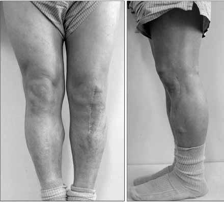
Fig. 6 Photographs, 14 months after corrective osteotomy, show normal alignment and correction of the genu recurvatum.
Reference
-
1. Chen LC, Chan YS, Wang CJ. Opening-wedge osteotomy, allografting with dual buttress plate fixation for severe genu recurvatum caused by partial growth arrest of the proximal tibial physis: a case report. J Orthop Trauma. 2004. 18:384–387.2. Coventry MB. Osteotomy about the knee for degenerative and rheumatoid arthiritis. J Bone Joint Surg Am. 1973. 55:23–48.3. Jackson JP. Osteotomy for osteoarthritis of the knee, in proceedings of the sheffield regional orthopedic club. J Bone Joint Surg. 1958. 40:826.4. Lee JY, Seon JK, Song EK, Yoon TR, Cheon SY, Lim KY. Comparison of high tibial osteotomy - opening versus closing wedge osteotomy -. J Korean Orthop Assoc. 2004. 39:790–796.5. Marti CB, Gautier E, Wachtl SW, Jakob RP. Accuracy of frontal and sagittal plane correction in open-wedge high tibial osteotomy. Arthroscopy. 2004. 20:366–372.
Article6. Moroni A, Pezzuto V, Pompili M, Zinghi G. Proximal osteotomy of the tibia for the treatment of genu recurvatum in adults. J Bone Joint Surg Am. 1992. 74:577–586.
Article7. Poilvache P. Scott WN, editor. Osteotomy for the arthritic knee: a European perspective. Surgery of the knee. 2006. 4th ed. Philadelphia: Elsevier Inc;1321–1366.8. Sundaram NA, Hallett JP, Sullivan MF. Dome osteotomy of the tibia for osteoarthritis of the knee. J Bone Joint Surg Br. 1986. 68:782–786.
Article9. Vail TP, Lang JE. Scott WN, editor. Surgical techniques and instrumentation in total knee arthroplasty. Surgery of the knee. 2006. 4th ed. Philadelphia: Elsevier Inc;1507–1508.
Article10. Winsor RE, Insall JN, Vince KG. Technical considerations of the total knee arthroplasty after proximal tibial osteotomy. J Bone Joint Surg Am. 1988. 70:547–555.
- Full Text Links
-
- Actions
-
Cited
- CITED
-
- Close
- Share
-
- Similar articles
-
- Delayed Onset of the Popliteal Artery Pseudoaneurysm Following Medial Opening Wedge High Tibial Osteotomy
- Interference of Union after the Use of Beta-Tricalcium Phosphate Block in High Tibial Osteotomy
- Treatment of genu recurvatum with the Ilizarov external fixator and proximal tibial corticotomy
- Ilizarov External Fixation in High Tibial Osteotomy
- Distal Femoral Medial Opening Wedge Osteotomy for Post-Traumatic, Distal Femoral Varus Deformity
