Inferior Accessory Ossicle of the Anterior Arch of the Atlas
- Affiliations
-
- 1Department of Orthopedic Surgery, College of Medicine, Dongguk University, Gyeongju, Korea. kjpil@dongguk.ac.kr
- 2Department of Radiology, College of Medicine, Dongguk University, Gyeongju, Korea.
- KMID: 2185524
- DOI: http://doi.org/10.4055/jkoa.2010.45.3.234
Abstract
- An inferior accessory ossicle of the anterior arch of the atlas is quite rare and should not be confused with other pathological conditions such as a fracture. Here we report a case of an inferior accessory ossicle of the anterior arch of the atlas in a 29-year-old male and a review of literature.
Keyword
Figure
-
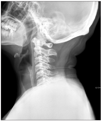
Figure 1 Lateral cervical radiograph shows loss of cervical lordotic curvature and bone fragment inferior to the anterior arch of the atlas. There is no prevertebral soft tissue swelling.
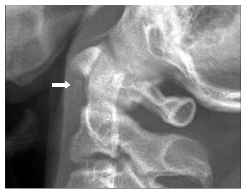
Figure 2 Magnified image of the upper cervical spine shows a well corticated inferior accessory ossicle of the anterior arch of the atlas (arrow).
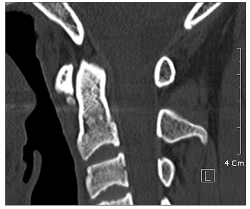
Figure 3 Sagittal CT image shows marginal sclerotic elliptical ossicle inferior to the anterior arch of the atlas.
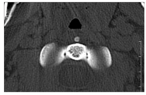
Figure 4 Axial CT image shows the midline location of the inferior accessory ossicle of the anterior arch of the atlas.
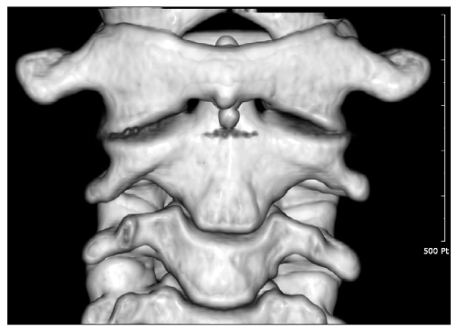
Figure 5 3D CT image viewed from anterior aspect of the upper cervical spine shows the inferior accessory ossicle of the anterior arch of the atlas.

Figure 6 (A) Flexion and (B) extension lateral cervical radiographs show no motion of the inferior accessory ossicle of the anterior arch of the atlas.
Reference
-
1. Mellado JM, Ramos A, Salvadó E, Camins A, Danús M, SaurÍ A. Accessory ossicles and sesamoid bones of the ankle and foot: imaging findings, clinical significance and differential diagnosis. Eur Radiol. 2003. 13:Suppl 4. L164–L177.
Article2. Keats TE. The inferior accessory ossicle of the anterior arch of the atlas. Am J Roentgenol Radium Ther Nucl Med. 1967. 101:834–836.
Article3. Naji MF, Bhat R. The typical appearance of the inferior accessory ossicle of the anterior arch of the atlas. Surg Radiol Anat. 2009. 31:69–71.
Article4. Von Lüdinghausen M, Fahr M, Prescher A, et al. Accessory joints between basiocciput and atlas/axis in the median plane. Clin Anat. 2005. 18:558–571.
Article5. Hall FM, Docken WP, Curtis HW. Calcific tendinitis of the longus coli: diagnosis by CT. Am J Roentgenol. 1986. 147:742–743.
Article6. Haun CL. Retropharyngeal tendinitis. AJR Am J Roentgenol. 1978. 130:1137–1140.
Article7. Jevtich V. Horizontal fracture of the anterior arch of the atlas. Case report. J Bone Joint Surg Am. 1986. 68:1094–1095.
Article8. Sasaka KK, Decker GT, El-Khoury GY. Horizontal fracture of the anterior arch of the atlas associated with a congenital cleft of the anterior arch. Emerg Radiol. 2006. 12:130–132.
Article
- Full Text Links
-
- Actions
-
Cited
- CITED
-
- Close
- Share
-
- Similar articles
-
- The Inferior Accessory Ossicle of the Anterior Arch of the Atlas Misdiagnosed as Anterior Arch Fracture: A Case Report
- Stress Fracture of the Anterior Atlas Arch Following C1 Posterior Arch Resection for Cervical Myelopathy with Retro-Odontoid Pseudotumor
- Spontaneous Anterior Atlas Fracture Following C1 Laminectomy without Fusion: A Case Report
- Hypertrophic Posterior Arch of Atlas Causing Cervical Myelopathy
- Congenital Anomaly of the Atlas Misdiagnosed as Posterior Arch Fracture of the Atlas and Atlantoaxial Subluxation
