J Korean Orthop Assoc.
2014 Aug;49(4):321-325. 10.4055/jkoa.2014.49.4.321.
Eighteen Year Follow-Up Results of Accessory Lower Limb Disarticulation and Pelvic Bone Reconstruction for Monocephalus Tripus Tribrachius
- Affiliations
-
- 1Department of Orthopaedic Surgery, Hanyang University College of Medicine, Seoul, Korea. sung1383@hanmail.net
- KMID: 2185196
- DOI: http://doi.org/10.4055/jkoa.2014.49.4.321
Abstract
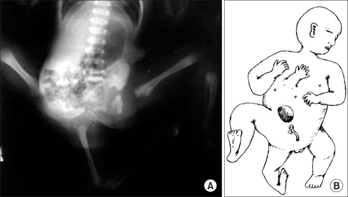
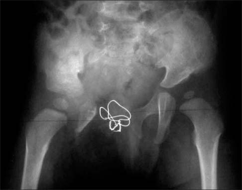
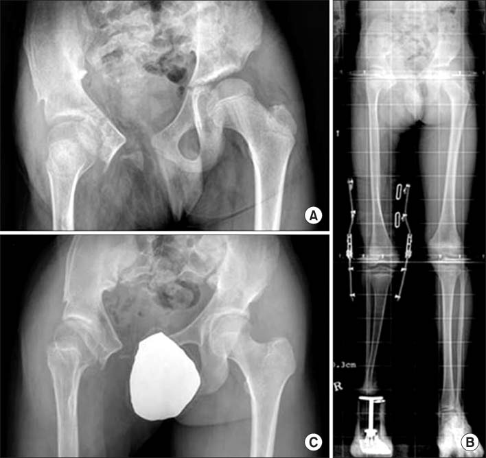
- Monocephalus tripus tribrachius, a type of conjoined twins with one head and three upper and lower extremities, is a rare congenital disorder. To date, no long-term follow-up results of surgical procedures for this condition have been reported in Korean literature. We experienced a case of monocephalus tripus tribrachius, which had been surgically managed with an accessory lower limb disarticulation and pelvic bone reconstruction to manage this accessory limb and accompanying comorbidities in hip joint and pelvis. Subsequently, ipsilateral Syme amputation was done for intractable deformity of foot, and later, ipsilateral femoral varus derotational osteotomy was done for inadequate coverage of femoral head observed in follow-up. We report 18-year follow-up results of the procedures with a review of literatures.
Keyword
MeSH Terms
Figure
Reference
-
1. O'Neill JA Jr, Holcomb GW 3rd, Schnaufer L, et al. Surgical experience with thirteen conjoined twins. Ann Surg. 1988; 208:299–312.2. Spencer R. Parasitic conjoined twins: external, internal (fetuses in fetu and teratomas), and detached (acardiacs). Clin Anat. 2001; 14:428–444.
Article3. Mahajan JK, Devendra K, Mainak D, Rao KL. Asymmetric conjoined twins: atypical ischiopagus parasite. J Pediatr Surg. 2002; 37:E33.
Article4. Corona-Rivera JR, Corona-Rivera E, Franco-Topete R, Acosta-León J, Aguila-Dueñas V, Corona-Rivera A. Atypical parasitic ischiopagus conjoined twins. . J Pediatr Surg. 2003; 38:e3.
Article5. Votteler TP, Lipsky K. Long-term results of 10 conjoined twin separations. J Pediatr Surg. 2005; 40:618–629.
Article6. Lee ES, Yoo CI. A case of "monocephalus tripus dibrachius". J Korean Orthop Assoc. 1971; 6:419–424.7. Gaine WJ, McCreath SW. Syme's amputation revisited: a review of 46 cases. J Bone Joint Surg Br. 1996; 78:461–467.8. Sponseller PD, Jani MM, Jeffs RD, Gearhart JP. Anterior innominate osteotomy in repair of bladder exstrophy. J Bone Joint Surg Am. 2001; 83:184–193.
Article9. Shultz WG. Plastic repair of exstrophy of bladder combined with bilateral osteotomy of ilia. J Urol. 1958; 79:453–458.
Article10. Vining NC, Song KM, Grady RW. Classic bladder exstrophy: orthopaedic surgical considerations. J Am Acad Orthop Surg. 2011; 19:518–526.
Article
- Full Text Links
- Actions
-
Cited
- CITED
-
- Close
- Share
- Similar articles
-
- Treatment of Large Arteriovenous Malformation in Right Lower Limb
- A Patient with Multiple Unfavorable Reconstruction Options: What Is the Best Choice?
- Fitting of a Myoelectric Hand for Wrist Disarticulation
- Recent Advances in Malignant Bone Tumor Treatment
- Comparative Analysis of Graft Patency and Limb Salvage Rate in DM & Non-DM after Infrainguinal Arterial Reconstruction


