An Extradural En-Plaque Meningioma Involving Thoracic Spine
- Affiliations
-
- 1Department of Orthopaedic Surgery, Dong-Eui Medical Center, Busan, Korea. wonro@hanmail.net
- 2Department of Pathology, Dong-Eui Medical Center, Busan, Korea.
- KMID: 2185178
- DOI: http://doi.org/10.4055/jkoa.2014.49.5.400
Abstract
- Reports of extradural spinal meningioma are rare, and differentiation from a metastatic lesion is important. We treated a case of thoracic extradural meningioma with surgical excision and obtained a favorable outcome without recurrence during one-year follow-up. Thus, we report on a case with review of the literature.
Keyword
MeSH Terms
Figure
-
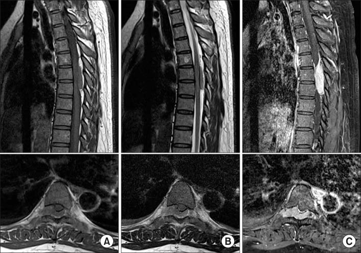
Figure 1 (A) On T1-weighted images, a mass separated from dura mater that compressed the spinal cord at the 6th and 7th thoracic vertebral level was seen on a sagittal image and a widened right neural foramen with compression of the 6th thoracic nerve root by the mass was seen on an axial image. (B) On T2-weighted images, the mass showed relatively homogeneous similar signal intensity with the spinal cord. (C) On gadolinium-enhanced images, the mass was enhanced homogeneously and distinguished from dura mater.
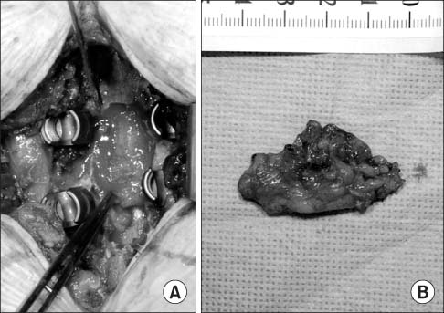
Figure 2 (A) The mass was seen on posterior dura mater intraoperatively. (B) The mass was completely excised.
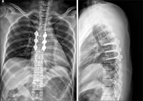
Figure 3 Postoperative plain radiograph showed posterior fixation.
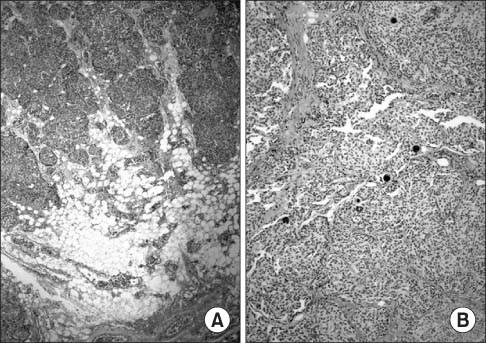
Figure 4 (A) The meningotheliomatous tumor cells were arranged in nests reminiscent of paraganglioma with hyalinized stromal vasculature. The tumor cells invaded into adjacent fat tissue (H&E, ×40). (B) Nests of meningotheliomatous tumor cells with several psammoma bodies and proliperated hyalinized vasculatures are noted. Mitosis is not seen (H&E, ×100).
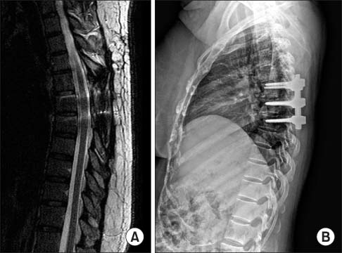
Figure 5 (A) On T2-weighted image, there was no local recurrence at one-year follow-up. (B) On a plain radiograph at one-year follow-up, mild kyphosis was seen.
Reference
-
1. Milz H, Hamer J. Extradural spinal meningiomas. Report of two cases. Neurochirurgia (Stuttg). 1983; 26:126–129.2. Fortuna A, Gambacorta D, Occhipinti EM. Spinal extradural meningiomas. Neurochirurgia (Stuttg). 1969; 12:166–180.
Article3. Sato N, Sze G. Extradural spinal meningioma: MRI. Neuroradiology. 1997; 39:450–452.
Article4. Haft H, Shenkin HA. Spinal epidural meningioma: case report. J Neurosurg. 1963; 20:801–804.5. Pecker MJ, Javalet A, Simon J, Loussouarn Y. Benign epidural tumors of the spinal cord. Neurochirurgie. 1967; 13:647–660.6. Stern J, Whelan MA, Correll JW. Spinal extradural meningiomas. Surg Neurol. 1980; 14:155–159.7. Kumar V, Abbas AK, Fausto N, Aster JC. Robbins & Cotran pathologic basis of disease. 8th ed. Philadelphia: Saunders;2010. p. 1338–1339.8. Mariniello G, Briganti F, De Caro ML, Maiuri F. Cervical extradural "en-plaque" meningioma. J Neurol Surg A Cent Eur Neurosurg. 2012; 73:330–333.
Article9. Barbanera A, Nina P, Serchi E, Ascanio F. Aggressive recurrence of intra-extradural cervico-thoracic meningothelial meningioma. Acta Neurochir (Wien). 2007; 149:83–86.
Article10. Solero CL, Fornari M, Giombini S, et al. Spinal meningiomas: review of 174 operated cases. Neurosurgery. 1989; 25:153–160.
Article
- Full Text Links
-
- Actions
-
Cited
- CITED
-
- Close
- Share
-
- Similar articles
-
- En Plaque Meningioma in Thoracic Spine: Case Report
- Extradural Spinal Lymphoplasmacyte-Rich Meningioma in the Thoracic Spine: A Case Report and Literature Review
- Convexity Meningioma En Plaque Presenting with Diffuse Hyperostosis of the Skull
- A Case Report of Meningioma "en plaque"
- Spinal Upper Thoracic Extradural Meningioma: A Case Report and Literature Review
