J Korean Orthop Assoc.  2014 Dec;49(6):422-430. 10.4055/jkoa.2014.49.6.422.

Sonographic Features of Common Soft Tissue Masses in the Extremities

Affiliations
  • 1Department of Radiology, Seoul National University College of Medicine, Seoul, Korea. dalnara3@gmail.com

Abstract

A wide variety of superficial soft tissue masses may be seen in clinical practice and imaging modalities can be helpful in achievement of a definitive diagnosis or limit a differential diagnosis. Among them, ultrasonography is well suited for screening soft tissue masses because of its safety, low cost, and real-time dynamic imaging. The purpose of this article is to review the characteristic sonographic appearance of soft tissue masses in the extremities.

Keyword

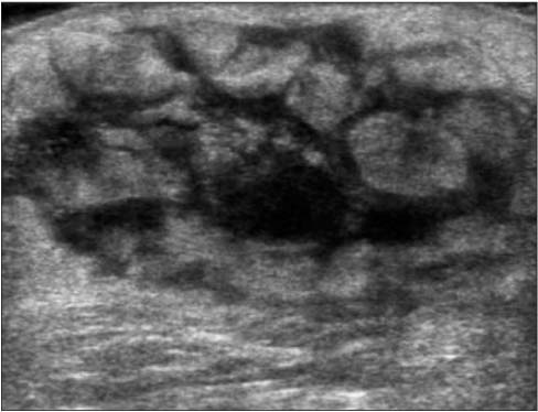
sarcoma; ultrasonography; soft tissue neoplasm

MeSH Terms

Diagnosis
Diagnosis, Differential
Extremities*
Mass Screening
Sarcoma
Soft Tissue Neoplasms
Ultrasonography*

Figure

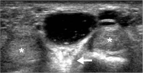
  • Figure 1 A 39-year-old female with ganglion between the 2nd and 3rd flexor tendons (asterisks) of the hand. Transverse sonographic image shows a well-circumscribed, anechoic mass with posterior acoustic enhancement (arrow).

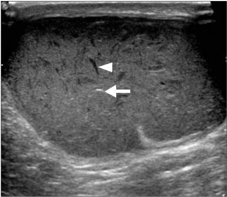
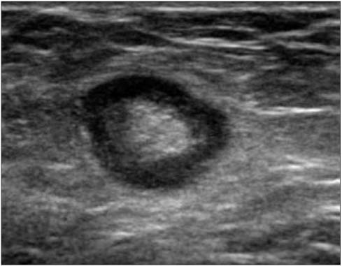
  • Figure 2 A 60-year-old male with an epidermal cyst of the upper arm. Transverse ultrasonography image shows a well-defined, solid, hyperechoic mass with posterior acoustic enhancement. The mass contains hyperechoic foci or linear echogenic reflectives (keratin, arrow) and a dark oil cleft (arrowhead).

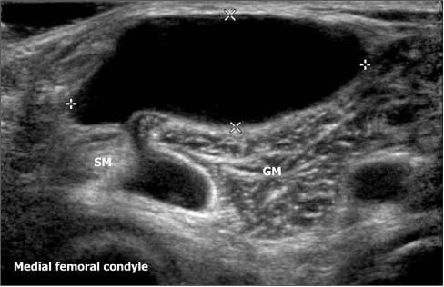
  • Figure 3 A 62-year-old male with a Baker cyst. Transverse ultrasonography image of popliteal fossa shows a cystic mass arising between tendons of the medial head of gastrocnemius (GM) and semimembranosus (SM) muscles.

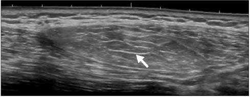
  • Figure 4 A 41-year-old female with subcutaneous lipoma of the back. Extended field-of-view ultrasonography image shows a large well-circumscribed, hyperechoic mass, characterized by thin and highly reflective linear echoes (arrow) oriented parallel to the skin, consistent with a lipoma.

  • Figure 5 A 32-year-old male with subcutaneous angiolipoma. (A) Transverse ultrasonography image shows a well-circumscribed, hyperechoic mass within the subcutaneous fat layer. (B) The Color Doppler image demonstrates vascularity within the mass, consistent with an angiolipoma.

  • Figure 6 A 28-year-old female with hemangioma. (A) Transverse ultrasonography image shows an ill-defined sponge-like mass containing hyperechoic dots with posterior acoustic shadowing, likely reflecting a phlebolith (arrows). (B) Corresponding Color Doppler image shows weak signals of flow within the soft tissue mass. (C) Radiographic correlation confirms the presence of a few phleboliths (arrow).

  • Figure 7 A 52-year-old female with venous malformation. The Color Doppler image shows an irregular hypoechoic mass with abnormal fluid-filled spaces and shows only a few, weak signals of flow within the soft tissue mass.

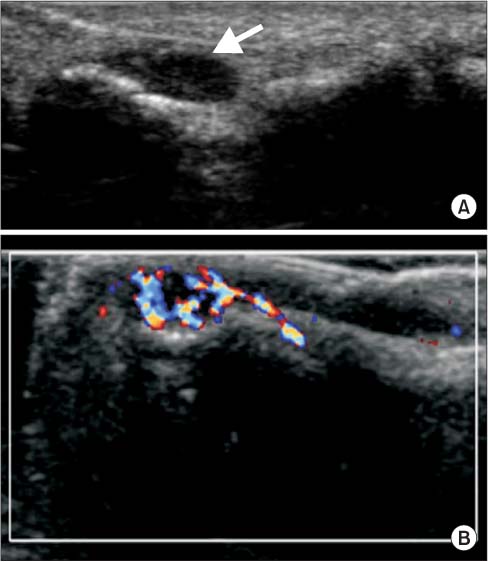
  • Figure 8 A 28-year-old female with a subungual glomus tumor in the finger tip. (A) Longitudinal ultrasonography (US) image shows a solid hypoechoic mass (arrow) beneath the nail. (B) Color Doppler US image shows marked intratumoral vasculature related to the high-velocity shunts of the glomus tumor.

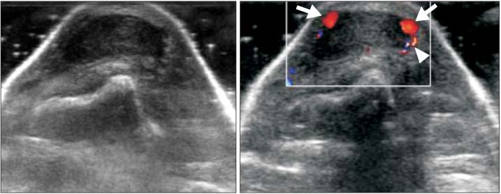
  • Figure 9 A 54-year-old female with a tenosynovial giant cell tumor in the finger. (A) Transverse ultrasonography (US) image shows a well-defined hypoehoic mass on the dorsal aspect of the extensor tendon. (B) Color Doppler US image shows displaced digital arteries (arrows) and sparse blood flow signals (arrowhead) within the tumor.

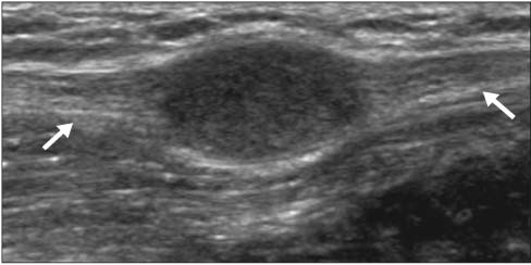
  • Figure 10 A 63-year-old male with a peripheral nerve sheath tumor. Longitudinal ultrasonography image shows a well-defined hypoehoic mass in continuity with the nerve (arrows).

  • Figure 11 A 65-year-old male with normal lymph node. Normal lymph node shows echogenic hilum.

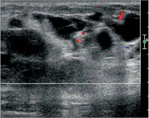
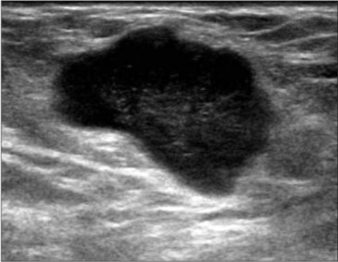
  • Figure 12 A 71-year-old male with a metastatic melanoma in the inguinal region. Transverse ultrasonography image shows an irregular marked hypoechoic mass without echogenic hilum.

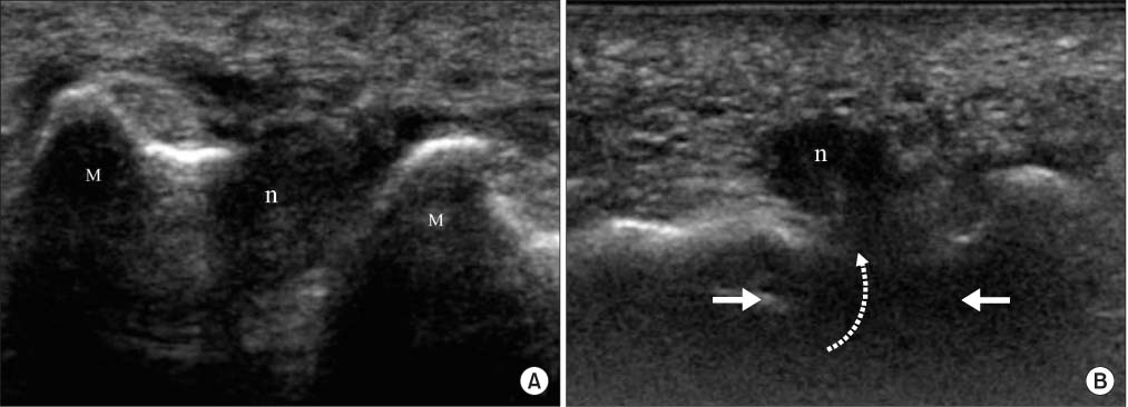
  • Figure 13 A 65-year-old female with Morton neuroma (n) in the third intermetatarsal space. (A) Transverse ultrasonography image shows an irregular marked hypoechoic mass in the third intermetatarsal space. (B) During the Mulder test, the increased lateral pressure (arrows) produces plantar displacement (dotted arrow) of the neuroma (n). This can be associated with a palpable 'click'. n, Morton neuroma; M, metatarsal bone.

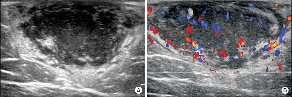
  • Figure 14 A 64-year-old male with dermatofibrosarcoma protuberans in the thigh. (A) Transverse ultrasonography image shows an irregular hypoechoic mass with an infiltrative margin, located in the subcutaneous layer. (B) Color Doppler image shows marked hypervascularity within the tumor.

  • Figure 15 A 64-year-old female with large B cell lymphoma. Transverse ultrasonography image shows a heterogeneous echoic mass with an ill-defined margin and intratumoral vascularity.


Reference

1. Lachenmayer A, Yang Q, Eisenberger CF, et al. Superficial soft tissue sarcomas of the extremities and trunk. World J Surg. 2009; 33:1641–1649.
Article
2. Kransdorf MJ. Imaging of soft tissue tumours. 2nd ed. Philadelphia: Lippincott Williams & Wilkins;2006.
3. Bianchi S, Martinoli C. Ultrasound of the musculoskeletal system. Berlin; New York: Springer;2007.
4. Lakkaraju A, Sinha R, Garikipati R, Edward S, Robinson P. Ultrasound for initial evaluation and triage of clinically suspicious soft-tissue masses. Clin Radiol. 2009; 64:615–621.
Article
5. Chiou HJ, Chou YH, Chiou SY, Wang HK. High-resolution ultrasonography in superficial soft tissue tumors. J Med Ultrasound. 2007; 15:152–174.
Article
6. Wu S, Tu R, Liu G, Shi Y. Role of ultrasound in the diagnosis of common soft tissue lesions of the limbs. Ultrasound Q. 2013; 29:67–71.
Article
7. Widmann G, Riedl A, Schoepf D, Glodny B, Peer S, Gruber H. State-of-the-art HR-US imaging findings of the most frequent musculoskeletal soft-tissue tumors. Skeletal Radiol. 2009; 38:637–649.
Article
8. Wu JS, Hochman MG. Soft-tissue tumors and tumorlike lesions: a systematic imaging approach. Radiology. 2009; 253:297–316.
Article
9. Teefey SA, Middleton WD, Boyer MI. Sonography of the hand and wrist. Semin Ultrasound CT MR. 2000; 21:192–204.
Article
10. Horcajadas AB, Lafuente JL, de la Cruz Burgos R, et al. Ultrasound and MR findings in tumor and tumor-like lesions of the fingers. Eur Radiol. 2003; 13:672–685.
Article
11. Dudea SM, Lenghel M, Botar-Jid C, Vasilescu D, Duma M. Ultrasonography of superficial lymph nodes: benign vs. malignant. Med Ultrason. 2012; 14:294–306.
12. Ahuja AT, Ying M. Sonographic evaluation of cervical lymph nodes. AJR Am J Roentgenol. 2005; 184:1691–1699.
Article
13. Chiou HJ, Chou YH, Chiu SY, et al. Differentiation of benign and malignant superficial soft-tissue masses using grayscale and color doppler ultrasonography. J Chin Med Assoc. 2009; 72:307–315.
Article
Full Text Links
  • JKOA
Actions
Cited
CITED
export Copy
Close
Share
  • Twitter
  • Facebook
Similar articles
Copyright © 2024 by Korean Association of Medical Journal Editors. All rights reserved.     E-mail: koreamed@kamje.or.kr