J Korean Fract Soc.
2016 Jan;29(1):68-72. 10.12671/jkfs.2016.29.1.68.
Calcified Anterior Tibial Artery Entrapment in Distal Third Tibial Fracture: A Case Report
- Affiliations
-
- 1Department of Orthopaedic Surgery, Yonsei University College of Medicine, Seoul, Korea.
- 2Department of Orthopaedic Surgery, College of Medicine, Konyang University, Daejeon, Korea. yougunwon@gmail.com
- KMID: 2183768
- DOI: http://doi.org/10.12671/jkfs.2016.29.1.68
Abstract
- In the distal third of the tibia, the anterior tibial artery runs close to the anterolateral surface of the tibial cortex. In a clinical situation, without vascular evaluation, injury or entrapment of the anterior tibial artery is difficult to detect. Because, an intact dorsalis pedis pulse is supplied with the collateral vessels of the posterior tibial artery. An entrapped anterior tibial artery can be injured during closed reduction in an emergency room or open reduction and internal fixation in the operating room. Care must be taken to prevent iatrogenic anterior tibial artery. In this case, an entrapped anterior tibial artery was observed in a simple radiograph and computed tomograph without contrast media for the vessel. We report on a rare case of calcified anterior tibial artery entrapment in a distal tibial fracture.
MeSH Terms
Figure
-

Fig. 1 An 80-year-old female patient with a spiral distal tibial fracture. (A) Antero-posterior view. (B) Lateral view. (C) Oblique view. Entrapped vessel is shown at the fracture site in the plain radiograph (arrow).
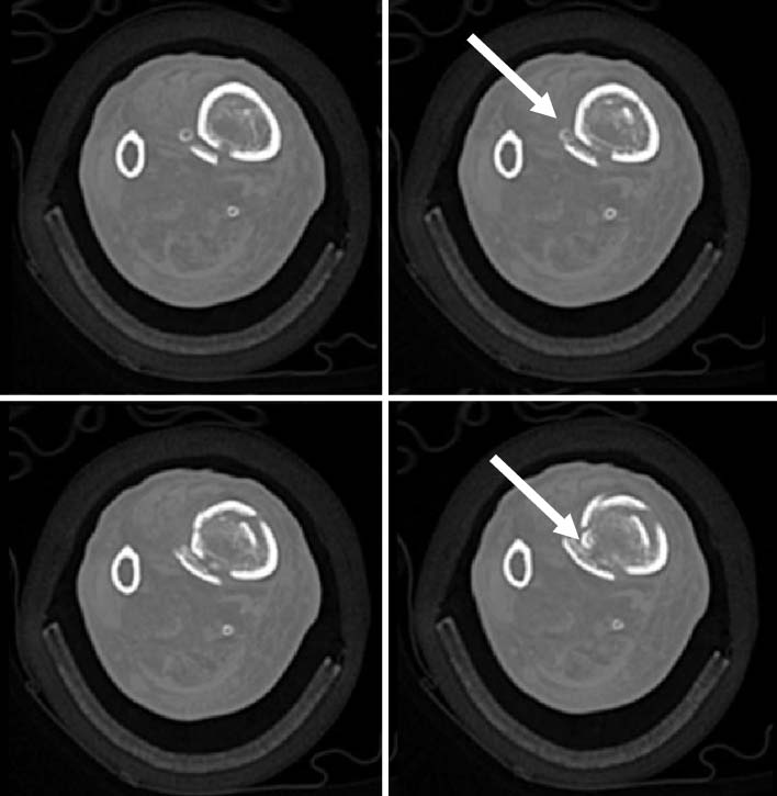
Fig. 2 Preoperative computed tomography axial images show the calcified anterior tibial artery (white arrows) at the fracture site.
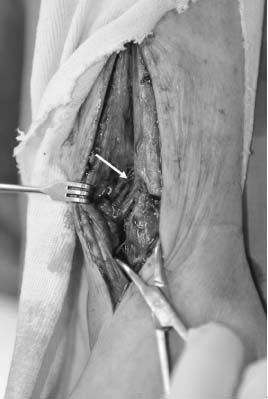
Fig. 3 Intraoperative photograph shows entrapment of the anterior tibial artery (arrow).
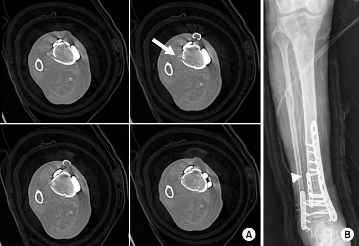
Fig. 4 (A) Postoperative computed tomography axial images show the reduction of calcified anterior tibial artery (arrow) in the fracture site and the fixation of fracture with locking compression plate. (B) Postoperative plain radiograph shows intact anterior tibial artery (arrow head).
Reference
-
1. Brinker MR, Bailey DE Jr. Fracture healing in tibia fractures with an associated vascular injury. J Trauma. 1997; 42:11–19.
Article2. Segal D, Brenner M, Gorczyca J. Tibial fractures with infrapopliteal arterial injuries. J Orthop Trauma. 1987; 1:160–169.
Article3. Tan ET, Tan TJ, Poon KB. Entrapment of the deep peroneal nerve and anterior tibial vessels by a spiral tibial fracture causing partial non-union: a case report. Skeletal Radiol. 2015; PMID: 26408316. [epub].
Article4. Court-Brown CM, McBirnie J. The epidemiology of tibial fractures. J Bone Joint Surg Br. 1995; 77:417–421.
Article5. Labler L, Wedler V, Mica L, Trentz O. Entrapment of the anterior tibial artery in a distal tibial fracture after intramedullary nailing. Unfallchirurg. 2006; 109:156–159.
Article6. Miki RA, Lawrence JP, Gillon TJ, Lawrence BD, Zell RA. Anterior tibial artery and deep peroneal nerve entrapment in spiral distal third tibia fracture. Orthopedics. 2008; 31:DOI: 10.3928/01477447-20081201-13. cited 2008 Dec. [Internet]. Available from: http://www.healio.com/orthopedics/trauma/journals/ortho/2008-12-31-12/%7B75123d28-e2b6-42a5-95b9-07ee2cb7b83f%7D/anterior-tibial-artery-and-deep-peroneal-nerve-entrapmentin-spiral-distal-third-tibia-fracture.
Article7. Sanders RJ, Alston GK. Variations and anomalies of the popliteal and tibial arteries. Am J Surg. 1986; 152:531–534.
Article8. Ebraheim NA, Lu J, Hao Y, Biyani A, Yeasting RA. Anterior tibial artery and its actual projection on the lateral aspect of the tibia: a cadaveric study. Surg Radiol Anat. 1998; 20:259–262.
Article9. Borrelli J Jr, Prickett W, Song E, Becker D, Ricci W. Extraosseous blood supply of the tibia and the effects of different plating techniques: a human cadaveric study. J Orthop Trauma. 2002; 16:691–695.
Article
- Full Text Links
-
- Actions
-
Cited
- CITED
-
- Close
- Share
-
- Similar articles
-
- Pseudoaneurysm of the Anterior Tibial Artery after Reduction with Pointed Bone Reduction Forceps on a Spiral Fracture of the Distal Tibia: A Case Report
- Pseudoaneurysm of the Anterior Tibial Artery After Closed Intramedullary Nailing of a Tibial Shaft Fracture: A Case Report
- Coil Embolization of a Pseudoaneurysm of the Anterior Tibial Artery: A Case Report
- Medullary Canal Widening Effect on Insertion of Tibial Intramedullary Nail bent anteriorly at distal portionJong Min Sohn
- Therapeutic Embolization for Pseudoaneurysm of the Anterior Tibial Artery after Tibial Nailing
