Surgical Treatment of Peroneus Longus Tendon Rupture after Ostectomy of Peroneus Tubercle of Calcaneus: A Case Report
- Affiliations
-
- 1Department of Orthopaedic Surgery, Kangdong Sacred Heart Hospital, Hallym University College of Medicine, Seoul, Korea. kiga9@hallym.or.kr
- KMID: 2181664
- DOI: http://doi.org/10.14193/jkfas.2014.18.2.72
Abstract
- We experienced a patient in whom rupture of the peroneus longus tendon occurred after ostectomy of the peroneus tubercle of the calcaneus. Acute rupture of the peroneus tendon can be managed by end-to-end anastomosis, while neglected cases can be treated by tenodesis, tendon transfer, or tendon graft. In the current patient, the tendon ends were mildly retracted, yielding a small gap. We successfully repaired the retracted tendon ends after lengthening by Z-plasty.
Figure
-

Figure 1. (A) Peroneus tubercle (white arrow) was seen in the three-dimensional reconstructed images of computed tomography performed before ostectomy of peroneus tubercle of calcaneus. (B) Irregular surface of peroneus tubercle stump (white arrow) was shown in the three-dimensional reconstructed images of computed tomography performed after ostectomy.

Figure 2. (A) Peroneus brevis tendon (white arrow) was found intact, but the continuity of peroneus longus tendon was not shown below the peroneus brevis tendon. (B) Distal stump of the ruptured peroneus longus tendon (white arrow) was found at the undersurface of the cuboid.
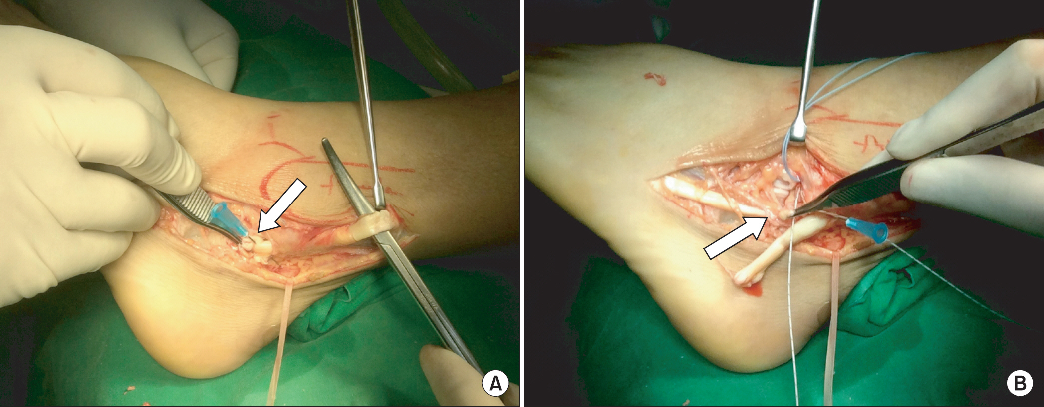
Figure 3. (A) Peroneus longus tendon (white arrow) was totally ruptured. (B) Peroneus brevis tendon and sural nerve (white arrow) were intact.

Figure 4. Peroneus longus tendon (white arrow) was repaired.

Figure 5. The continuity of repaired peroneus longus tendon (white arrow) was confirmed below the peroneus brevis tendon (arrowhead) in the coronal plane of the postoperative magnetic resonance imaging.
Reference
-
References
1. Arbab D, Tingart M, Frank D, Abbara-Czardybon M, Waizy H, Wingenfeld C. Treatment of isolated peroneus longus tears and a review of the literature. Foot Ankle Spec. 2014; 7:113–8.
Article2. Ng VY, Hothem E, Calhoun J. Radiologic case study. Peroneus longus rupture. Orthopedics. 2010; 33(701):778–9.3. Dombek MF, Lamm BM, Saltrick K, Mendicino RW, Catanzariti AR. Peroneal tendon tears: a retrospective review. J Foot Ankle Surg. 2003; 42:250–8.
Article4. Clarke HD, Kitaoka HB, Ehman RL. Peroneal tendon injuries. Foot Ankle Int. 1998; 19:280–8.
Article5. Redfern D, Myerson M. The management of concomitant tears of the peroneus longus and brevis tendons. Foot Ankle Int. 2004; 25:695–707.
Article6. Krause JO, Brodsky JW. Peroneus brevis tendon tears: pathophysiology, surgical reconstruction, and clinical results. Foot Ankle Int. 1998; 19:271–9.
Article7. Brandes CB, Smith RW. Characterization of patients with primary peroneus longus tendinopathy: a review of twenty-two cases. Foot Ankle Int. 2000; 21:462–8.
Article8. Heckman DS, Reddy S, Pedowitz D, Wapner KL, Parekh SG. Operative treatment for peroneal tendon disorders. J Bone Joint Surg Am. 2008; 90:404–18.
Article
- Full Text Links
-
- Actions
-
Cited
- CITED
-
- Close
- Share
-
- Similar articles
-
- Operative Treatment of Stenosing Tenosynovitis of the Peroneus Longus Tendon Associated with Hypertrophy of the Peroneal Tubercle: A Case Report - 1 Case
- Fracture of the Os Peroneum with Rupture of the Peroneus Longus Tendon: A Case Report
- Sural Nerve Entrapment and Tenosynovitis of Peroneus Longus by Hypertrophied Peroneal Tubercle: A Case Report
- Total Rupture of Peroneus Longus Tendon Through an Os Peroneum Fracture Treated by Tendon Transfer (A Case Report)
- Fracture of Os Peroneum with Rupture of the Peroneus Longus Tendon: A Case Report
