Complications of Scarf Osteotomy for Hallux Valgus
- Affiliations
-
- 1Department of Orthopedic Surgery, Pohang St. Mary's Hospital, Pohang, Korea. scarpel@gmail.com
- KMID: 2181638
- DOI: http://doi.org/10.14193/jkfas.2014.18.4.178
Abstract
- PURPOSE
The purpose of this study was to evaluate the frequency of troughing and stress fracture, which are the major complications of scarf osteotomy, and to suggest methods to prevent these complications.
MATERIALS AND METHODS
We reviewed 243 cases of 137 patients treated with the scarf osteotomy for hallux valgus from January 2005 to December 2012. The mean follow-up period was 2.8 years. During the scarf osteotomy, a long oblique longitudinal osteotomy was performed in order to decrease the possibility of troughing and stress fracture. Radiographs of lateral view of the foot were obtained and the thicknesses of the first metatarsal base at the sagittal plane were measured and compared.
RESULTS
There was no troughing during fragment translation and screw fixation intraoperatively. Radiographs of lateral view of the foot taken preoperatively and at the last follow-up showed that the mean thickness of the first metatarsal was 22.4 mm preoperatively and 21.6 mm at the last follow-up, with a mean difference of 0.8 mm. And no stress fracture was observed.
CONCLUSION
To prevent troughing and stress fracture, a long oblique longitudinal cut, parallel to the first metatarsal plantar surface, was performed, making both ends of the proximal segment truncated cone-shape, and securing the strong bony strut of the proximal segment. No troughing or stress fracture was experienced with scarf osteotomy.
Keyword
Figure
-
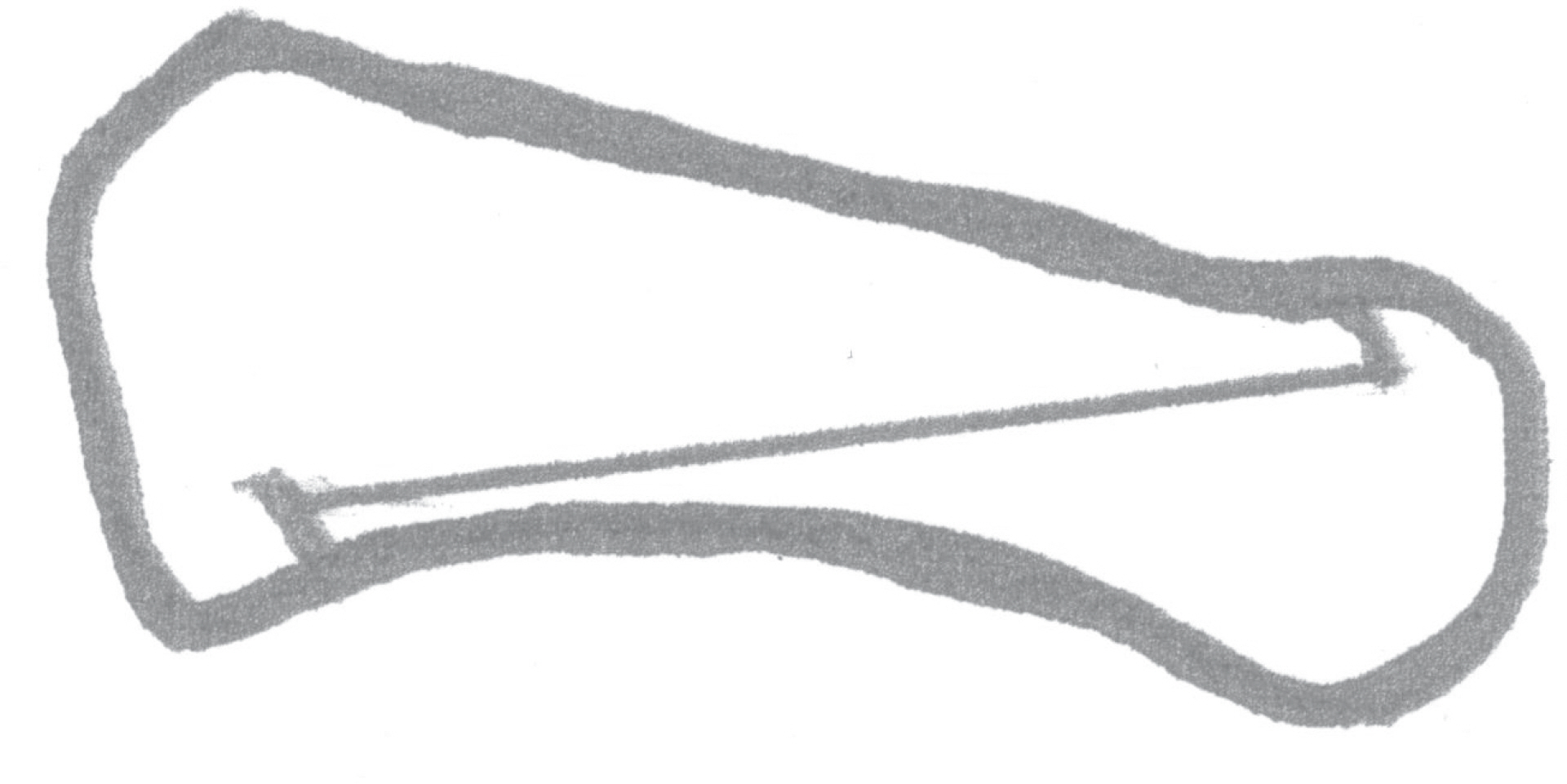
Figure 1. This picture shows longitudinal and transverse cut line in scarf osteotomy. Longitudinal cut is made from proximal plantar to distal dorsal area of the first metatarsal.
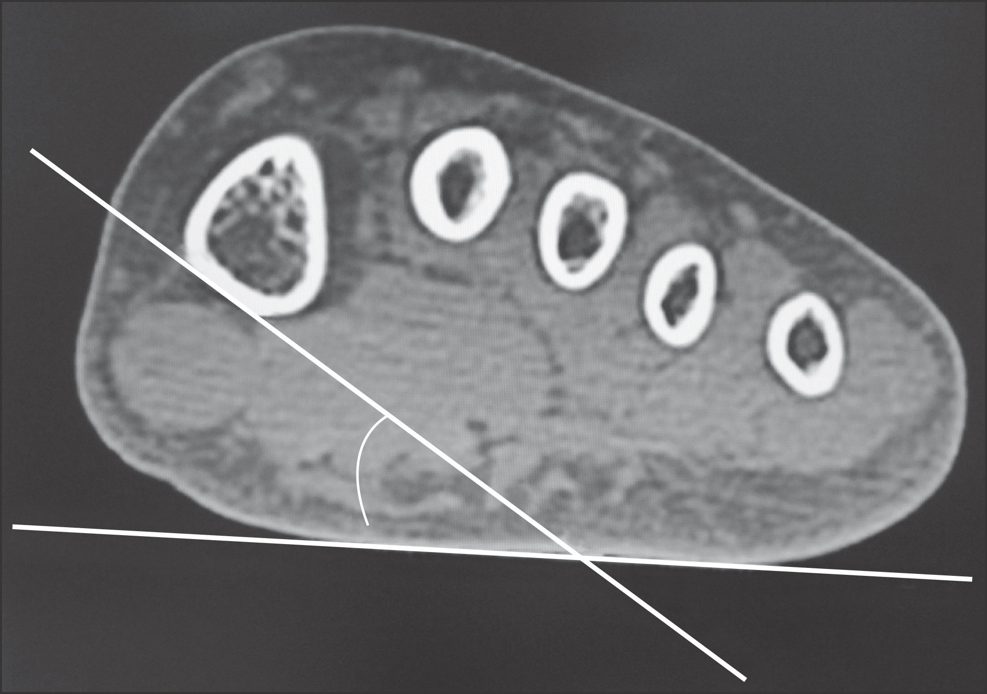
Figure 2. This is an axial plane image of computed tomography of a left foot taken 1∼2 cm distal to the metatarsal base. Note the angle between the plantar surface of first metatarsal and the plantar surface of foot sole.
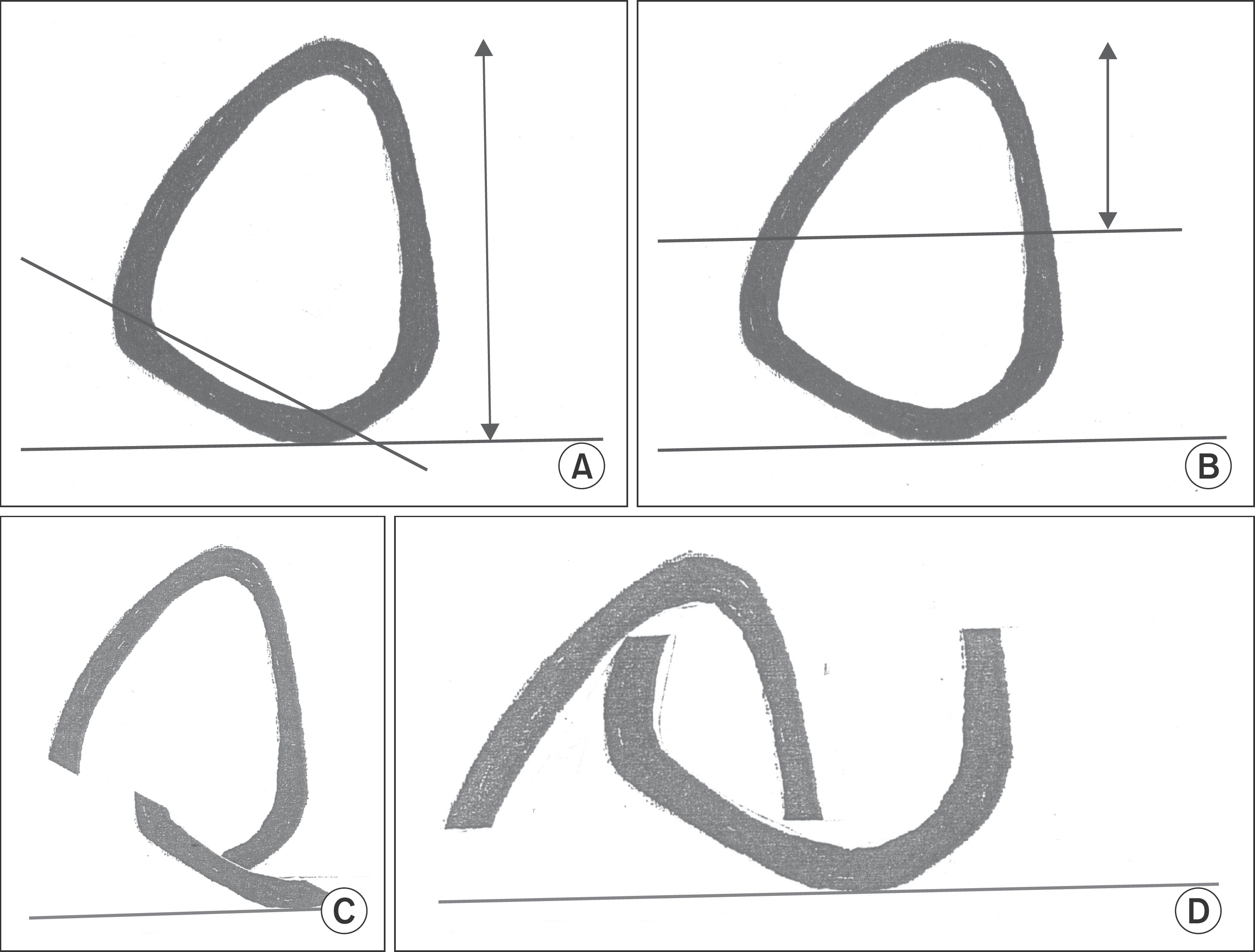
Figure 3. These pictures show two different longitudinal cut crossing the 1st metatarsal transversally and after translation at 1∼2 cm distal to the metatarsal base. (A) Cut line is parallel to the plantar surface of first metatarsal bone. This direction of cut line makes lateral strut of dorsal fragment wide and strong (double headed arrow, lateral surface). (B) Cut line is not parallel to the plantar surface of 1st metatarsal bone. This cut results in narrow lateral strut and semi-circular dorsal fragment in coronal plane (double headed arrow, lateral surface). (C) During translation of each fragment, there is little chance of troughing, because of its structural character. (D) Also, during translation, the chance of troughing could be increased because of its relatively narrow and semi-circular lateral surface of first metatarsal bone.
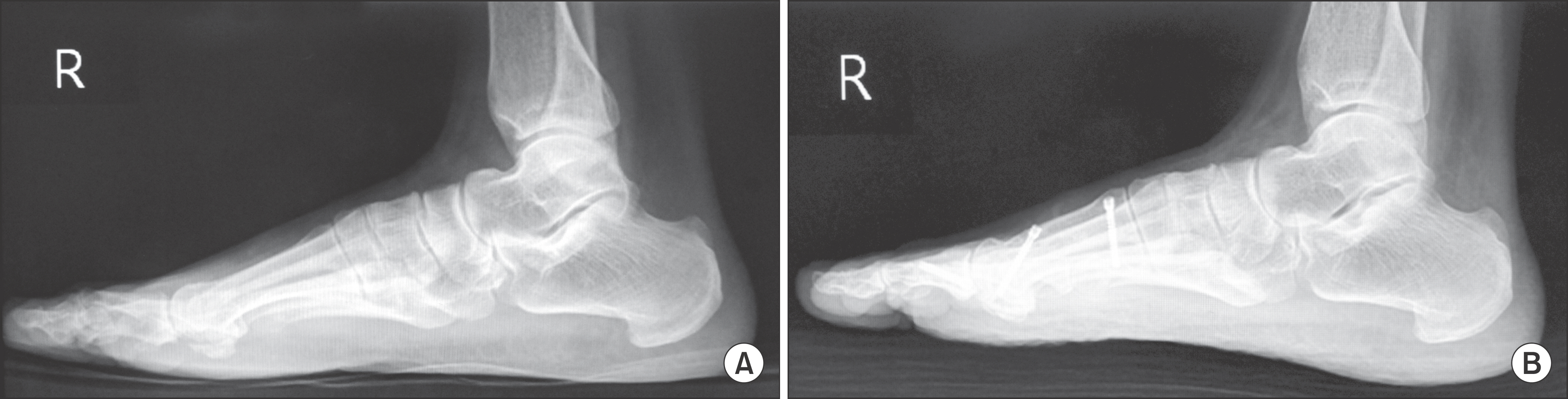
Figure 4. (A) This is a preoperative lateral radiograph of a right foot before scarf osteotomy. (B) At the last follow-up after scarf osteotomy, there were no remarkable changes in the thickness of first metatarsal.
Cited by 2 articles
-
Comparative Analysis of the Results between the Early Period and the Midterm Period of a Single Surgeon’s Experience in the Treatment of Hallux Valgus Using Scarf Osteotomy
Yeong-Hyeon Lee, Il-Hyun Nam, Tae-Hun Lee, Gil-Yeong Ahn, Yong-Sik Lee, Sung-Hyun Hwang, Kyung-Jin Lee
J Korean Foot Ankle Soc. 2020;24(4):135-141. doi: 10.14193/jkfas.2020.24.4.135.Rotational Long Scarf Osteotomy on Hallux Valgus in Elderly Patients with Osteoporosis
Il Hyun Nam, Dae Geun Kim, Young Hoon Lee, Dong Hyun Lee, Young Duk Choi, Hee Hyung Lee
J Korean Orthop Assoc. 2018;53(5):415-420. doi: 10.4055/jkoa.2018.53.5.415.
Reference
-
1.Chung JW., Jung HW., Chu IT. Modified scarf osteotomy for hallux valgus with lesser metatarsalgia. J Korean Foot Ankle Soc. 2008. 12:134–9.2.Park HW., Kim SJ. Treatment results of hallux valgus deformity by parallel-shaped modified scarf osteotomy. J Korean Foot Ankle Soc. 2012. 16:123–7.3.Kristen KH., Berger C., Stelzig S., Thalhammer E., Posch M., Engel A. The SCARF osteotomy for the correction of hallux valgus deformities. Foot Ankle Int. 2002. 23:221–9.
Article4.Schneider W., Csepan R., Knahr K. Reproducibility of the radiographic metatarsophalangeal angle in hallux surgery. J Bone Joint Surg Am. 2003. 85:494–9.
Article5.Smith AM., Alwan T., Davies MS. Perioperative complications of the Scarf osteotomy. Foot Ankle Int. 2003. 24:222–7.
Article6.Kramer J., Barry LD., Helfman DN., Mehnert JA., Pokrifcak VM. The modified Scarf bunionectomy. J Foot Surg. 1992. 31:360–7.7.Barouk LS. Scarf osteotomy for hallux valgus correction. Local anatomy, surgical technique, and combination with other forefoot procedures. Foot Ankle Clin. 2000. 5:525–58.8.Jones S., Al Hussainy HA., Ali F., Betts RP., Flowers MJ. Scarf oste-otomy for hallux valgus. A prospective clinical and pedobaro-graphic study. J Bone Joint Surg Br. 2004. 86:830–6.9.Coetzee JC. Scarf osteotomy for hallux valgus repair: the dark side. Foot Ankle Int. 2003. 24:29–33.
Article10.Steck JK., Ringstrom JB. Long Z-osteotomy: a review and new modification to correct troughing. J Foot Ankle Surg. 2001. 40:305–10.
Article11.Weil LS. Scarf osteotomy for correction of hallux valgus. Historical perspective, surgical technique, and results. Foot Ankle Clin. 2000. 5:559–80.12.Zygmunt KH., Gudas CJ., Laros GS. Z-bunionectomy with internal screw fixation. J Am Podiatr Med Assoc. 1989. 79:322–9.
Article13.Schoen NS., Zygmunt K., Gudas C. Z-bunionectomy: retrospective long-term study. J Foot Ankle Surg. 1996. 35:312–7.
Article14.Duke HF. Rotational scarf (Z) osteotomy bunionectomy for cor-rection of high intermetatarsal angles. J Am Podiatr Med Assoc. 1992. 82:352–60.
Article15.Miller JM., Stuck R., Sartori M., Patwardhan A., Cane R., Vrbos L. The inverted Z bunionectomy: quantitative analysis of the scarf and inverted scarf bunionectomy osteotomies in fresh cadaveric matched pair specimens. J Foot Ankle Surg. 1994. 33:455–62.16.Crevoisier X., Mouhsine E., Ortolano V., Udin B., Dutoit M. The scarf osteotomy for the treatment of hallux valgus deformity: a review of 84 cases. Foot Ankle Int. 2001. 22:970–6.
Article17.Berg RP., Olsthoorn PG., Pöll RG. Scarf osteotomy in hallux valgus: a review of 72 cases. Acta Orthop Belg. 2007. 73:219–23.18.Barouk LS. Drawbacks with this type of hallux valgus surgery. Barouk LS, editor. editor.Forefoot reconstruction. 2nd ed.Paris, New York: Springer;2005. p. 94–105.
- Full Text Links
-
- Actions
-
Cited
- CITED
-
- Close
- Share
-
- Similar articles
-
- Treatment Results of Hallux Valgus Deformity by Parallel-Shaped Modified Scarf Osteotomy
- Scarf Osteotomy for the Treatment of Recurred Hallux Valgus
- Short Scarf Osteotomy for Moderate Hallux Valgus
- Modified Proximal Scarf Osteotomy for Hallux Valgus
- The Effect of Derotational Closing Wedge Akin Osteotomy for the Treatment of Hallux Valgus with the Pronation of Great Toe
