Prosthetic rehabilitation for a patient with CO-MI discrepancy
- Affiliations
-
- 1Department of Prosthodontics and Research Institute of Oral Science College of Dentistry, Gangneung-Wonju National University, Gangneung, Republic of Korea. doctorcj@gwnu.ac.kr
- KMID: 2180030
- DOI: http://doi.org/10.14368/jdras.2015.31.3.273
Abstract
- Centric occlusion-maximum intercuspation (CO-MI) discrepancy is one of main causes of evoking premature contact and resultant mandibular shift. These non-physiological conditions can induce temporomandibular disease, periodontitis, and non-carious cervical lesion. Therefore, if CO-MI discrepancy exists in patients who need extensive prosthetic rehabilitation, it must be corrected and then physiological occlusion must be restored. This report describes the treatment procedure of removing CO-MI discrepancy and prosthetic rehabilitation in a patient with 3.5 mm discrepancy, multiple caries and periodontitis. Proper mandibular position and modified opening & closing movement were confirmed by ARCUSdigma II and transcranial radiograph.
Figure
-

Fig. 1 Radiographic evaluation: severe bone loss and secondary caries on upper left quadrant.
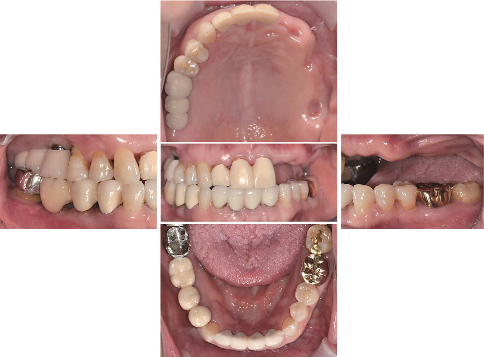
Fig. 2 Intraoral examination: multiple teeth loss, gingival recession and midline discrepancy.
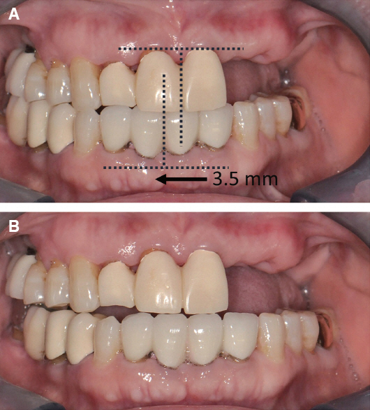
Fig. 3 (A) right mandibular shift on maximum intercuspation, (B) guided centric relation.
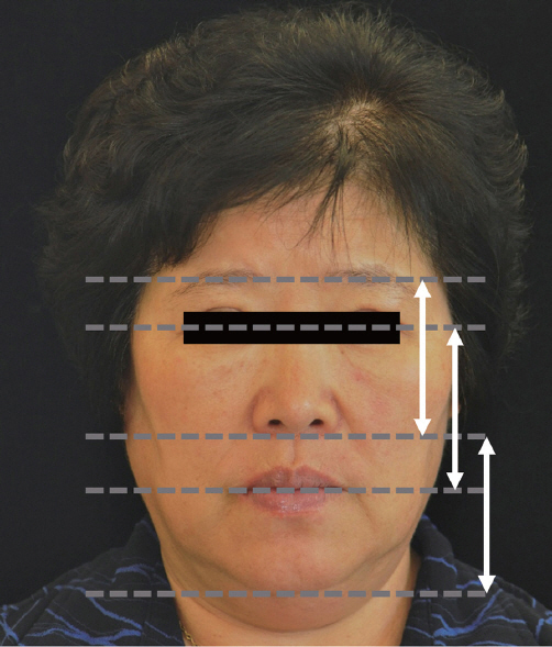
Fig. 4 Vertical dimension evaluation.
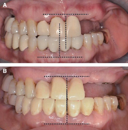
Fig. 5 (A) initial examination, (B) corrected mandibular position using 1st provisional prosthesis.
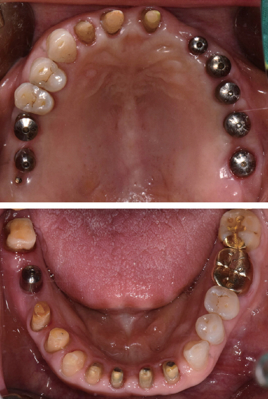
Fig. 6 Re-evaluation for prosthetic and occlusal design after implant placement and teeth preparation.
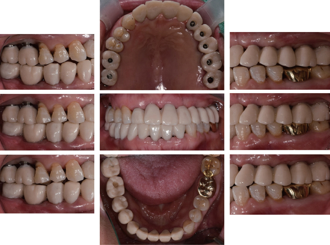
Fig. 7 Stable centric and lateral occlusal contact establishment using 2nd provisional prosthesis.
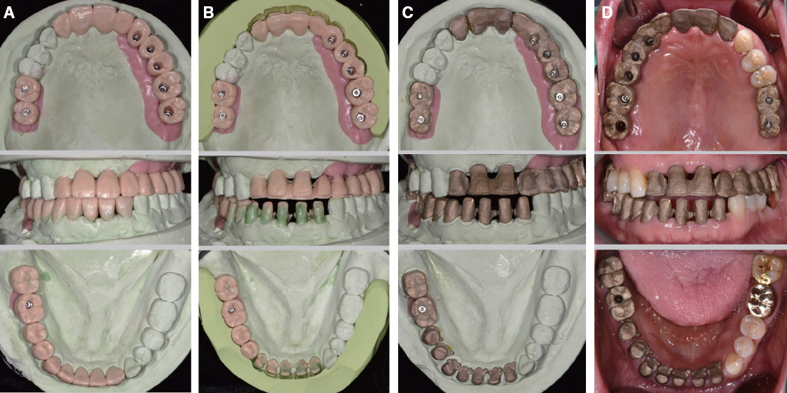
Fig. 8 (A) Full-contour wax-up, (B) Cut-back, (C) Metal coping fabrication, (D) Try-in.

Fig. 9 Definitive prosthesis showing harmonized occlusal relationship.
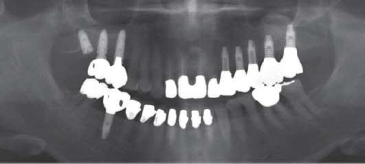
Fig. 10 Panoramic radiograph at 3 month follow-up.

Fig. 11 Transcranial view of right mandibular condyle. (A) At initial examination, (B) During provisionalization, (C) After Wearing definitive prosthesis.
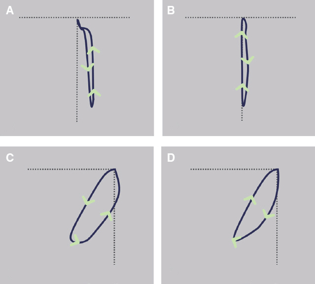
Fig. 12 Opening and closing path traced by ARCUSdigma II (A) Frontal view on initial examination, (B) Frontal view after definitive prosthesis delivery, (C) Sagittal view on initial examination, (D) Sagittal view after definitive prosthesis delivery.
Reference
-
References
1. The glossary of prosthodontic terms. J Prosthet Dent. 2005; 94:10–92. DOI: 10.1016/j.prosdent.2005.03.013.2. Pullinger AG, Seligman DA, Gornbein JA. A multiple logistic regression analysis of the risk and relative odds of temporomandibular disorders as a function of common occlusal features. J Dent Res. 1993; 72:968–79. DOI: 10.1177/00220345930720061301. PMID: 8496480.3. Landi N, Manfredini D, Tognini F, Romagnoli M, Bosco M. Quantification of the relative risk of multiple occlusal variables for muscle disorders of the stomatognathic system. J Prosthet Dent. 2004; 92:190–5. DOI: 10.1016/j.prosdent.2004.05.013. PMID: 15295330.4. Branschofsky M, Beikler T, Schäfer R, Flemming TF, Lang H. Secondary trauma from occlusion and periodontitis. Quintessence Int. 2011; 42:515–22. PMID: 21519589.5. Harrel SK, Nunn ME. The association of occlusal contacts with the presence of increased periodontal probing depth. J Clin Periodontol. 2009; 36:1035–42. DOI: 10.1111/j.1600-051X.2009.01486.x. PMID: 19930093.6. Brandini DA, Trevisan CL, Panzarini SR, Pedrini D. Clinical evaluation of the association between noncarious cervical lesions and occlusal forces. J Prosthet Dent. 2012; 108:298–303. DOI: 10.1016/S0022-3913(12)60180-2.7. Troeltzsch M, Troeltzsch M, Cronin RJ, Brodine AH, Frankenberger R, Messlinger K. Prevalence and association of headaches, temporomandibular joint disorders, and occlusal interferences. J Prosthet Dent. 2011; 105:410–7. DOI: 10.1016/S0022-3913(11)60084-X.8. Dawson PE. Functional occlusion: from TMJ to smile design. 2007. St. Louis: CV Mosby;p. 4–9. PMID: 17906220.9. Ehrlich J, Hochman N, Yaffe A. The masticatory pattern as an adjunct for diagnosis and treatment. J Oral Rehabil. 1992; 19:393–8. DOI: 10.1111/j.1365-2842.1992.tb01581.x. PMID: 1432354.10. Yoshida E, Fueki K, Igarashi Y. Association between food mixing ability and mandibular movements during chewing of a wax cube. J Oral Rehabil. 2007; 34:791–9. DOI: 10.1111/j.1365-2842.2007.01743.x. PMID: 17919244.11. Park JM, Kim HJ, Park EJ, Kim MR, Kim SJ. Three dimensional finite element analysis of the stress distribution around the mandibular posterior implant during non-working movement according to the amount of cantilever. J Adv Prosthodont. 2014; 6:361–71. DOI: 10.4047/jap.2014.6.5.361. PMID: 25352958. PMCID: PMC4211052.12. Aglietta M, Siciliano VI, Zwahlen M, Brägger U, Pjetursson BE, Lang NP, Salvi GE. A systematic review of the survival and complication rates of implant supported fixed dental prostheses with cantilever extensions after an observation period of at least 5 years. Clin Oral Implants Res. 2009; 20:44151. DOI: 10.1111/j.1600-0501.2009.01706.x. PMID: 19522975.13. Zurdo J, Romão C, Wennström JL. Survival and complication rates of implant-supported fixed partial dentures with cantilevers: a systematic review. Clin Oral Implants Res. 2009; 20(Suppl4):59–66. DOI: 10.1111/j.1600-0501.2009.01773.x. PMID: 19663951.14. Salama MA, Salama H, Garber DA. Guidelines for aesthetic restorative options and implant site enhancement: the utilization of orthodontic extrusion. Pract Proced Aesthet Dent. 2002; 14:125–130.15. Hellsing G. Occlusal adjustment and occlusal stability. J Prosthet Dent. 1988; 59:696–702. DOI: 10.1016/0022-3913(88)90385-X.16. Kirveskari P. Assessment of occlusal stability by measuring contact time and centric slide. J Oral Rehabil. 1999; 26:763–6. DOI: 10.1046/j.1365-2842.1999.00483.x. PMID: 10564430.
- Full Text Links
-
- Actions
-
Cited
- CITED
-
- Close
- Share
-
- Similar articles
-
- Rehabilitation of a patient with non-syndromic partial oligodontia
- Rehabilitation of the Bilateral Upper Extremity Amputees
- Prosthetic ambulation in a paraplegic patient with a transfemoral amputation and radial nerve palsy
- Prosthetic Gait Training in Individuals with Pathologic Conditions and Associated Pain on the Non-Amputated Side
- Prosthetic rehabilitation for a glossectomy patient: a clinical report
