J Clin Neurol.
2013 Apr;9(2):103-110. 10.3988/jcn.2013.9.2.103.
Juxtacortical Spots on Fluid-Attenuated Inversion Recovery Images in Cryptogenic Transient Ischemic Attack
- Affiliations
-
- 1Department of Neurology, Cerebrovascular Center, Chonnam National University Medical School, Gwangju, Korea. alldelight2@jnu.ac.kr
- 2Research Institute of Medical Sciences, Chonnam National University, Gwangju, Korea.
- 3Department of Neurology, Chonnam National University Hwasun Hospital, Hwasun, Korea.
- KMID: 2179169
- DOI: http://doi.org/10.3988/jcn.2013.9.2.103
Abstract
- BACKGROUND AND PURPOSE
Juxtacortical spots are detected frequently on fluid-attenuated inversion recovery (FLAIR) images, but have not been extensively researched in patients with transient ischemic attack (TIA). We hypothesized that juxtacortical spots on FLAIR images are partly associated with right-to-left shunt (RLS) in TIA without clear etiology. The possibility of an association between the presence of RLS and juxtacortical spots on FLAIR images in patients with TIA without clear etiology was investigated, and the imaging findings of patients with and without RLS were compared.
METHODS
This was a retrospective study of TIA patients who visited our tertiary stroke center consecutively within 72 hours of TIA onset. Cryptogenic TIA was defined as no clear etiology despite a routine diagnostic workup. The presence of RLS was examined by transcranial Doppler with an agitated saline test or transesophageal echocardiography. Juxtacortical spots were defined as small and round hyperintensities in the juxtacortex on FLAIR images, excluding white-matter hyperintensities.
RESULTS
Of the 132 patients with cryptogenic TIA examined for this study, 70 (53.0%) had RLS. Juxtacortical spots on FLAIR images were detected more frequently in patients with RLS than in those without. The independent factors for the presence of juxtacortical spots were RLS [odds ratio (OR)=3.802, 95% confidence interval (95% CI)=1.74-8.2; p=0.001] and age (OR=1.058, 95% CI=1.01-1.10; p=0.004) by multivariate analysis. The number of juxtacortical spots was significantly higher among patients with a moderate-to-large RLS than in those with a small or no RLS.
CONCLUSIONS
The findings of the present study demonstrate a significant association between the presence of RLS and the occurrence of juxtacortical spots on FLAIR images in patients with cryptogenic TIA.
Keyword
MeSH Terms
Figure
-
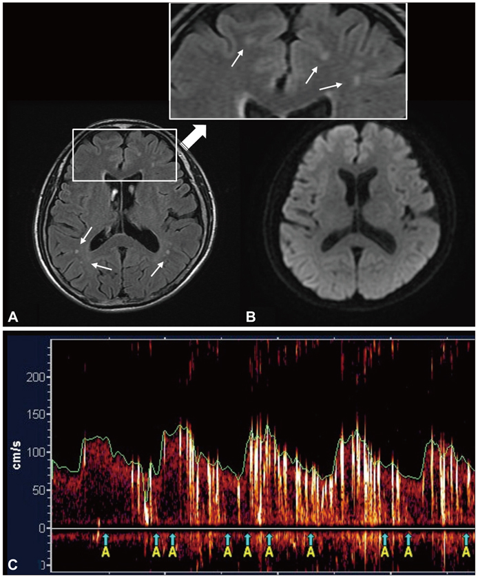
Fig. 1 Imaging findings of a 45-year-old woman with transient left hemiparesis. (A) Juxtacortical spots on FLAIR images (arrows). (B) Normal DWI. (C) Multiple high-intensity transient signals during contrast TCD monitoring of the MCA. DWI: diffusion-weighted imaging, FLAIR: fluid-attenuated inversion recovery, TCD: transcranial Doppler.
Reference
-
1. Special report from the National Institute of Neurological Disorders and Stroke. Classification of cerebrovascular diseases III. Stroke. 1990. 21:637–676.2. Albers GW, Caplan LR, Easton JD, Fayad PB, Mohr JP, Saver JL, et al. Transient ischemic attack--proposal for a new definition. N Engl J Med. 2002. 347:1713–1716.
Article3. Warach S, Kidwell CS. The redefinition of TIA: the uses and limitations of DWI in acute ischemic cerebrovascular syndromes. Neurology. 2004. 62:359–360.
Article4. Kidwell CS, Alger JR, Di Salle F, Starkman S, Villablanca P, Bentson J, et al. Diffusion MRI in patients with transient ischemic attacks. Stroke. 1999. 30:1174–1180.
Article5. Crisostomo RA, Garcia MM, Tong DC. Detection of diffusion-weighted MRI abnormalities in patients with transient ischemic attack: correlation with clinical characteristics. Stroke. 2003. 34:932–937.
Article6. Ay H, Oliveira-Filho J, Buonanno FS, Schaefer PW, Furie KL, Chang YC, et al. 'Footprints' of transient ischemic attacks: a diffusion-weighted MRI study. Cerebrovasc Dis. 2002. 14:177–186.
Article7. Inatomi Y, Kimura K, Yonehara T, Fujioka S, Uchino M. DWI abnormalities and clinical characteristics in TIA patients. Neurology. 2004. 62:376–380.
Article8. Coutts SB, Simon JE, Eliasziw M, Sohn CH, Hill MD, Barber PA, et al. Triaging transient ischemic attack and minor stroke patients using acute magnetic resonance imaging. Ann Neurol. 2005. 57:848–854.
Article9. Handke M, Harloff A, Olschewski M, Hetzel A, Geibel A. Patent foramen ovale and cryptogenic stroke in older patients. N Engl J Med. 2007. 357:2262–2268.
Article10. Lamy C, Giannesini C, Zuber M, Arquizan C, Meder JF, Trystram D, et al. Clinical and imaging findings in cryptogenic stroke patients with and without patent foramen ovale: the PFO-ASA Study. Atrial Septal Aneurysm. Stroke. 2002. 33:706–711.
Article11. Steiner MM, Di Tullio MR, Rundek T, Gan R, Chen X, Liguori C, et al. Patent foramen ovale size and embolic brain imaging findings among patients with ischemic stroke. Stroke. 1998. 29:944–948.
Article12. Johnston SC, Rothwell PM, Nguyen-Huynh MN, Giles MF, Elkins JS, Bernstein AL, et al. Validation and refinement of scores to predict very early stroke risk after transient ischaemic attack. Lancet. 2007. 369:283–292.
Article13. Fazekas F, Kleinert R, Offenbacher H, Schmidt R, Kleinert G, Payer F, et al. Pathologic correlates of incidental MRI white matter signal hyperintensities. Neurology. 1993. 43:1683–1689.
Article14. Jauss M, Zanette E. Detection of right-to-left shunt with ultrasound contrast agent and transcranial Doppler sonography. Cerebrovasc Dis. 2000. 10:490–496.
Article15. Hamann GF, Schätzer-Klotz D, Fröhlig G, Strittmatter M, Jost V, Berg G, et al. Femoral injection of echo contrast medium may increase the sensitivity of testing for a patent foramen ovale. Neurology. 1998. 50:1423–1428.
Article16. Droste DW, Silling K, Stypmann J, Grude M, Kemény V, Wichter T, et al. Contrast transcranial doppler ultrasound in the detection of right-to-left shunts: time window and threshold in microbubble numbers. Stroke. 2000. 31:1640–1645.
Article17. Clergeau MR, Hamon M, Morello R, Saloux E, Viader F, Hamon M. Silent cerebral infarcts in patients with pulmonary embolism and a patent foramen ovale: a prospective diffusion-weighted MRI study. Stroke. 2009. 40:3758–3762.
Article18. Kovacs IB, Gorog DA, Yamamoto J. Enhanced spontaneous thrombolysis: a new therapeutic challenge. J Thromb Thrombolysis. 2006. 21:221–227.
Article19. Ueno Y, Shimada Y, Tanaka R, Miyamoto N, Tanaka Y, Hattori N, et al. Patent foramen ovale with atrial septal aneurysm may contribute to white matter lesions in stroke patients. Cerebrovasc Dis. 2010. 30:15–22.
Article20. Yoon GJ, Kim JT, Chang J, Kim DE, Cho BH, Lee JH, et al. Right-to-left shunts as a cause of juxtacortical spots in patients with migraine. Eur J Neurol. 2012. 19:1086–1092.
Article
- Full Text Links
-
- Actions
-
Cited
- CITED
-
- Close
- Share
-
- Similar articles
-
- Reversible Cerebral Vasoconstriction Syndrome Presenting as Transient Vessel Wall Enhancement on Contrast-Enhanced Fluid-Attenuated Inversion Recovery Images: A Case Report and Literature Review
- Concomitant Small Intracerebral Hemorrhage in a Patient with Cerebral Amyloid Angiopathy Mimicking Transient Ischemic Attack
- Selective Gray Matter Infarction in the Basal Ganglia Associated With Transient Ischemic Attack
- Usefulness of Combined Fat- and Fluid-Suppressed SPIR-FLAIR Images in Optic Neuritis: Comparison with Fat-Suppressed SPIR or STIR Images or STIR images
- Hyperintensity of Subarachnoid Space on FLAIR Images Caused by Supplemental Oxygen
